
 中文
中文 English
 한국어
한국어 日本語
日本語 Español
 Français
 Русский
| Catalog No. | HW776023 | ||||
|---|---|---|---|---|---|
| Species reactivity | Human | ||||
| Applications | ELISA, FCM, IP | ||||
| Host species | Mouse | ||||
| Clonality | Monoclonal | ||||
| Isotype | IgG1, kappa | ||||
| Clone ID | 3D1 | ||||
| Tested applications | WB, FCM | ||||
| Target | T-lymphocyte activation antigen CD86, Activation B7-2 antigen, B70, BU63, CTLA-4 counter-receptor B7.2, FUN-1, CD86, CD86, CD28LG2, B7-2 | ||||
| Endotoxin level | Please contact with the lab for this information. | ||||
| Purity | >95% as determined by SDS-PAGE. | ||||
| Purification | Protein A/G purified from cell culture supernatant. | ||||
| Accession | P42081, K7GMU1 | ||||
| Form | Liquid | ||||
| Storage buffer | 0.01M PBS pH 7.4 Please refer to the specific buffer information in the hardcopy of datasheet or the lot-specific COA. |
||||
| Reconstitution | Reconstitute in sterile water for a stock solution. A copy of datasheet will be provided with the products, please refer to it for details. | ||||
| Product Usage Information |
| ||||
| Stability and Storage | Use a manual defrost freezer and avoid repeated freeze thaw cycles. Store at 2 to 8°C for frequent use. Store at -20 to -80°C for twelve months from the date of receipt. | ||||
| Background | T-lymphocyte activation antigen CD86 (B7-2) is a ~37 kDa protein. Costimulatory molecule that belongs to the immunoglobulin superfamily that plays an important role in T-lymphocyte activation. Acts as the primary auxiliary signal augmenting the MHC/TCR signal in naive T-cells by acting as a ligand for the CD28 receptor which is constitutively expressed on the cell surface of T-cells. May play a critical role in the early events of T-cell activation and costimulation of naive T-cells, such as deciding between immunity and anergy that is made by T-cells within 24 hours after activation. Also involved in the regulation of B cells function, plays a role in regulating the level of IgG(1) produced. Upon CD40 engagement, activates NF-kappa-B signaling pathway via phospholipase C and protein kinase C activation. 1. Collins, AV. et al. (2002) Immunity 17, 201-10. PMID: 12196291 2. Freeman, GJ. et al. (1993) Science (New York, N.Y.) 262, 909-11. PMID: 7694363 3. Lanier, LL. et al. (1995) Journal of immunology (Baltimore, Md. : 1950) 154, 97-105. PMID: 7527824 4. Schwartz, JC. et al. (2001) Nature 410, 604-8. PMID: 11279501 | ||||
| Note | For research use only | ||||
| Isotype Control | Mouse IgG1, kappa Recombinant Isotype Control Antibody (MOPC-21) (MV080207) |
Western blot analysis was performed using anti-CD86/B7-2 monoclonal antibody at 1ug/mL on various samples.
Lane 1: recombinant human CD86/B7-2 (Catalog No: HW776011)
Lane 2: negative control
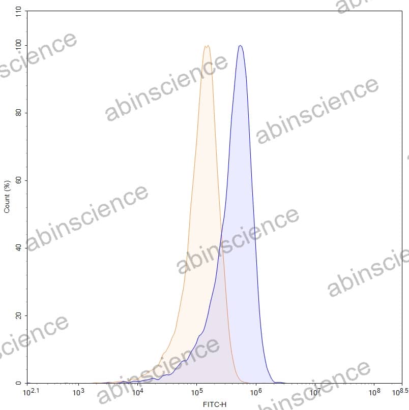
Flow-cytometry using anti-human CD86 antibody. Daudi cells were stained with an irrelevant antibody (Blue Histogram) or an anti-human CD86 monoclonal antibody (Catalog HW776023, Yellow Histogram) at a concentration of 5 µg/ml for 30 mins at RT. After washing, bound antibody was detected using a Goat Anti-Mouse IgG H&L Polyclonal Antibody, FITC (abinScience: MF690414) and cells analysed on a NovoCyte Flow Cytometer.
+86-27-65523339
Building C, No. 666, Shen Dun Si Lu, Wuhan, 430206, China


