
 中文
中文 English
 한국어
한국어 日本語
日本語 Español
 Français
 Русский


| Catalog No. | HB167023 | ||||
|---|---|---|---|---|---|
| Species reactivity | Human | ||||
| Applications | ELISA, FCM | ||||
| Host species | Mouse | ||||
| Clonality | Monoclonal | ||||
| Isotype | IgG2a, kappa | ||||
| Clone ID | M3/38 | ||||
| Target | Mac-2 antigen, Lectin L-29, LGALS3, Galactose-specific lectin 3, 35 kDa lectin, Galactoside-binding protein, CBP 35, L-31, Gal-3, Laminin-binding protein, Galectin-3, GALBP, IgE-binding protein, Carbohydrate-binding protein 35, MAC2 | ||||
| Endotoxin level | Please contact with the lab for this information. | ||||
| Purity | >95% as determined by SDS-PAGE. | ||||
| Purification | Protein A/G purified from cell culture supernatant. | ||||
| Accession | P17931 | ||||
| RRID | Anti-Human Gal3/Galectin-3 Antibody (M3/38) (abinScience Cat# HB167023, RRID:AB_3729067) |
||||
| Form | Liquid | ||||
| Storage buffer | 0.01M PBS pH 7.4 Please refer to the specific buffer information in the hardcopy of datasheet or the lot-specific COA. |
||||
| Product Usage Information |
| ||||
| Stability and Storage | Use a manual defrost freezer and avoid repeated freeze-thaw cycles. Store at 4°C short term (1-2 weeks). Store at -20°C 12 months. Store at -80°C long term. | ||||
| Background | Galectin-3 (Gal-3) is a ~26 kDa protein. Galactose-specific lectin which binds IgE. May mediate with the alpha-3, beta-1 integrin the stimulation by CSPG4 of endothelial cells migration. Together with DMBT1, required for terminal differentiation of columnar epithelial cells during early embryogenesis. In the nucleus: acts as a pre-mRNA splicing factor. Involved in acute inflammatory responses including neutrophil activation and adhesion, chemoattraction of monocytes macrophages, opsonization of apoptotic neutrophils, and activation of mast cells. | ||||
| Note | For research use only. | ||||
| Isotype Control | Mouse IgG2a (Fc Silence) Recombinant Isotype Control (MOPC 173) (MF533023) |
Detects Human LGALS3 in indirect ELISAs.
Flow-cytometry using anti-human Galectin-3 antibody.HaCaT cells were stained with an irrelevant antibody (Blue Histogram) or an anti-human Galectin-3 antibody monoclonal antibody (Catalog # HB167023 ,Green Histogram) at a concentration of 5 µg/ml for 30 mins at RT. After washing, bound antibody was detected using a FITC conjugated goat anti-mouse antibody (Catalog # MF690414) and cells analysed on a NovoCyte Flow Cytometer.
Flow-cytometry using anti-human Galectin-3 antibody.MCF7 cells were stained with an irrelevant antibody (Blue Histogram) or an anti-human Galectin-3 antibody monoclonal antibody (Catalog # HB167023 ,Green Histogram) at a concentration of 5 µg/ml for 30 mins at RT. After washing, bound antibody was detected using a FITC conjugated goat anti-mouse antibody (Catalog # MF690414) and cells analysed on a NovoCyte Flow Cytometer.
Flow-cytometry using anti-human Galectin-3 antibody.HeLa cells were stained with an irrelevant antibody (Blue Histogram) or an anti-human Galectin-3 antibody monoclonal antibody (Catalog # HB167023 ,Green Histogram) at a concentration of 5 µg/ml for 30 mins at RT. After washing, bound antibody was detected using a FITC conjugated goat anti-mouse antibody (Catalog # MF690414) and cells analysed on a NovoCyte Flow Cytometer.
Flow-cytometry using anti-human Galectin-3 antibody.SiHa cells were stained with an irrelevant antibody (Blue Histogram) or an anti-human Galectin-3 antibody monoclonal antibody (Catalog # HB167023 ,Green Histogram) at a concentration of 5 µg/ml for 30 mins at RT. After washing, bound antibody was detected using a FITC conjugated goat anti-mouse antibody (Catalog # MF690414) and cells analysed on a NovoCyte Flow Cytometer.
SDS-PAGE for Anti-Human Gal3/Galectin-3 Antibody (M3/38).
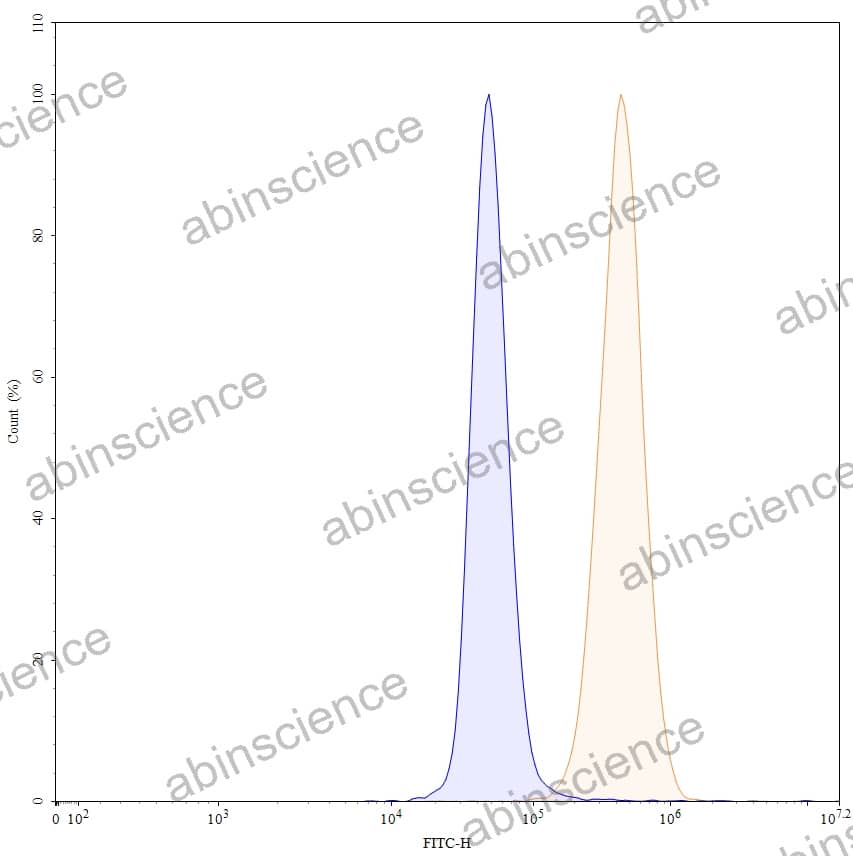
Flow-cytometry using anti-human Galectin-3 antibody. 5637 cells were fixed and permeabilized, then stained with an irrelevant antibody (Blue Histogram) or an anti-human Galectin-3 monoclonal antibody (Catalog HB167023, Yellow Histogram) at a concentration of 5 µg/ml for 30 mins at RT. After washing, bound antibody was detected using Goat Anti-Mouse IgG H&L Polyclonal Antibody, FITC (abinScience: MF690414) and cells analysed on a NovoCyte Flow Cytometer.


Contact us for custom quotes, bulk requests and any other issues.
Mail: [email protected]
+86-27-65523339
Building C, No. 666, Shen Dun Si Lu, Wuhan, 430206, China


