
 中文
中文 English
 한국어
한국어 日本語
日本語 Español
 Français
 Русский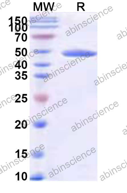

| Catalog No. | HB833014 | ||||||||
|---|---|---|---|---|---|---|---|---|---|
| Description |
Anti-Human TUBG1 Polyclonal Antibody (HB833014) is a rabbit polyclonal antibody detecting TUBG1 in ELISA, IHC, WB. Suitable for Human.
Highlights
|
||||||||
| Species reactivity | Human, Mouse, Rat | ||||||||
| Applications | ELISA, IHC, WB | ||||||||
| Host species | Rabbit | ||||||||
| Clonality | Polyclonal | ||||||||
| Isotype | IgG | ||||||||
| Immunogen | E. coli - derived recombinant Human TUBG1 (Met1-Gln451). | ||||||||
| Target | Tubulin gamma-1 chain, Gamma-1-tubulin, Gamma-tubulin complex component 1, GCP-1, TUBG1, TUBG | ||||||||
| Purification | Purified by antigen affinity column. | ||||||||
| Accession | P23258 | ||||||||
| Form | Liquid | ||||||||
| Storage buffer | 0.01M PBS, pH 7.4, 50% Glycerol, 0.05% Proclin 300. Please refer to the specific buffer information in the hardcopy of datasheet or the lot-specific COA. |
||||||||
| Product Usage Information |
| ||||||||
| Stability and Storage | Use a manual defrost freezer and avoid repeated freeze thaw cycles. Store at 2 to 8°C for frequent use. Store at -20 to -80°C for twelve months from the date of receipt. | ||||||||
| Background | Tubulin gamma-1 chain (TUBG1) is a ~51 kDa protein. Tubulin is the major constituent of microtubules, protein filaments consisting of alpha- and beta-tubulin heterodimers. Gamma-tubulin is a key component of the gamma-tubulin ring complex (gTuRC) which mediates microtubule nucleation. The gTuRC regulates the minus-end nucleation of alpha-beta tubulin heterodimers that grow into microtubule protafilaments, a critical step in centrosome duplication and spindle formation. 1. Brito, C. et al. (2024) Science (New York, N.Y.) 383, 870-876. PMID: 38305685 2. Aher, A. et al. (2024) Nature structural & molecular biology 31, 1124-1133. PMID: 38609661 3. Serna, M. et al. (2024) Developmental cell 59, 3175-3188.e8. PMID: 39321809 | ||||||||
| Note | For research use only. |
Western blot analysis was performed using anti-TUBG1 polyclonal antibody at 1ug/mL on various samples.
Lane 1: NIH3T3 cell lysate
Lane 2: HepG2 cell lysate
Lane 3: K562 cell lysate
Lane 4: Hela cell lysate
Lane 5: Jurkat cell lysate
Lane 6: A431 cell lysate
Lane 7: C2C12 cell lysate
Lane 8: U2OS cell lysate
Lane 9: A549 cell lysate
Contact us for custom quotes, bulk requests and any other issues.
Mail: [email protected]
+86-27-65523339
Building C, No. 666, Shen Dun Si Lu, Wuhan, 430206, China


