
 中文
中文 English
 한국어
한국어 日本語
日本語 Español
 Français
 Русский
| Catalog No. | MX128014 | ||||||||
|---|---|---|---|---|---|---|---|---|---|
| Description |
Anti-Mouse PPIA Polyclonal Antibody (MX128014) is a rabbit polyclonal antibody detecting Ppia. Suitable for Rat, Mouse, and Human.
Highlights
|
||||||||
| Species reactivity | Human, Mouse, Rat | ||||||||
| Applications | ELISA, IHC, WB | ||||||||
| Host species | Rabbit | ||||||||
| Isotype | IgG | ||||||||
| Clonality | Polyclonal | ||||||||
| Immunogen | E. coli - derived recombinant Mouse PPIA (Met1-Leu164). | ||||||||
| Target | Cyclophilin A, Cyclosporin A-binding protein, EC:5.2.1.8, PPIase A, Peptidyl-prolyl cis-trans isomerase A, Peptidyl-prolyl cis-trans isomerase A, N-terminally processed, Ppia, Rotamase A, SP18 | ||||||||
| Endotoxin level | Please contact with the lab for this information. | ||||||||
| Purification | Purified by antigen affinity column. | ||||||||
| Accession | P17742 | ||||||||
| Form | Liquid | ||||||||
| Storage buffer | 0.01M PBS, pH 7.4, 50% Glycerol, 0.05% Proclin 300. Please refer to the specific buffer information in the hardcopy of datasheet or the lot-specific COA. |
||||||||
| Product Usage Information |
| ||||||||
| Background | Peptidyl-prolyl cis-trans isomerase A (PPIA) is a ~17 kDa protein. Catalyzes the cis-trans isomerization of proline imidic peptide bonds in oligopeptides. Exerts a strong chemotactic effect on leukocytes partly through activation of one of its membrane receptors BSG/CD147, initiating a signaling cascade that culminates in MAPK/ERK activation. Activates endothelial cells (ECs) in a pro-inflammatory manner by stimulating activation of NF-kappa-B and ERK, JNK and p38 MAP-kinases and by inducing expression of adhesion molecules including SELE and VCAM1. Induces apoptosis in ECs by promoting the FOXO1-dependent expression of CCL2 and BCL2L11 which are involved in EC chemotaxis and apoptosis. In response to oxidative stress, initiates proapoptotic and antiapoptotic signaling in ECs via activation of NF-kappa-B and AKT1 and up-regulation of antiapoptotic protein BCL2. 1. Wang, L. et al. (2014) Thrombosis and haemostasis 111, 873-82. PMID: 24429998 2. Lauranzano, E. et al. (2015) Brain : a journal of neurology 138, 974-91. PMID: 25678563 | ||||||||
| Note | For research use only |
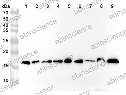
Western blot analysis was performed using anti-PPIA polyclonal antibody at 1ug/mL on various samples.
Lane 1: HepG2 cell lysate
Lane 2: NIH3T3 cell lysate
Lane 3: K562 cell lysate
Lane 4: Raji cell lysate
Lane 5: Jurkat cell lysate
Lane 6: PC-12 cell lysate
Lane 7: 293T cell lysate
Lane 8: Mouse brain lysate
Lane 9: Rat brain lysate
Contact us for custom quotes, bulk requests and any other issues.
Mail: [email protected]
+86-27-65523339
Building C, No. 666, Shen Dun Si Lu, Wuhan, 430206, China


