I. Overview of Vasculitis: The Immune System's "Faulty Orders" and Vascular Damage
Vasculitis refers to a group of inflammatory disorders characterized by immune system dysfunction, leading to aberrant immune attacks on the body's own blood vessel walls. This pathological assault results in inflammatory infiltration, thickening, and increased fragility of the vessel walls, which can progress to lumen narrowing, occlusion, and in severe cases, vessel rupture and hemorrhage. Ultimately, impaired blood perfusion to target organs leads to functional damage.
Given the ubiquitous distribution of blood vessels throughout the body, the clinical manifestations of vasculitis are highly heterogeneous. Often termed "the great mimicker," it can be easily confused with infectious diseases, dermatological conditions, renal diseases, and others. Clinical symptoms can be divided into systemic and local manifestations. Systemic symptoms commonly include fever, fatigue, and weight loss. Local symptoms correspond to the target organs supplied by the affected vessels and may include skin purpura, arthralgia, cough with hemoptysis, hematuria, and limb numbness. The precise etiology of vasculitis remains incompletely understood but is thought to involve genetic predisposition, infectious triggers, drug exposure, among other factors. Most types follow a chronic, progressive course, making early diagnosis and standardized treatment crucial for improving patient prognosis.
II. Classification of Vasculitis: A Disease Spectrum Based on Vessel Size
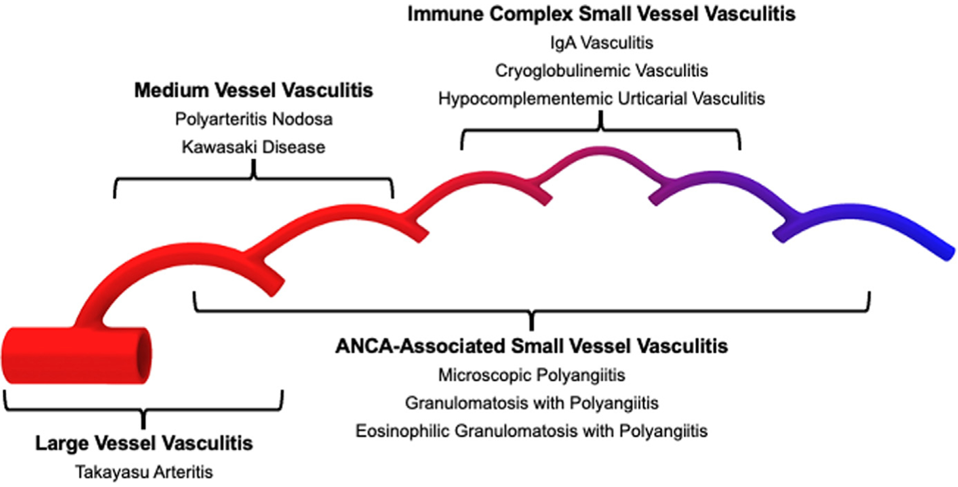
Figure 1. Distribution of vessel involvement by large vessel vasculitis (LVV), medium vessel vasculitis (MVV), and small vessel vasculitis (SVV) (Doi: 10.1016/j.rdc.2021.07.007)
The classification system for vasculitis is complex. The 2012 Chapel Hill Consensus Conference (CHCC) classification criteria are widely adopted in clinical practice. These criteria are centered on the size of the predominantly affected blood vessels, combined with clinical features and pathological findings, categorizing vasculitis into three main classes, each with characteristic representative diseases:
1. Large Vessel Vasculitis (LVV)
Primarily affects the aorta and its major branches. The disease course is chronic and progressive, often leading to vascular stenosis, occlusion, or aneurysm formation. Common types include:
- Giant Cell Arteritis (GCA): Predominantly occurs in individuals over 50 years old, with higher incidence in Northern European populations and a female predominance. It mainly involves cranial arteries and can lead to irreversible vision loss or stroke if severe.
- Takayasu Arteritis (TAK): More common in women under 40, with higher prevalence in Asian populations. It can involve the entire aorta and its branches, clinically presenting with limb claudication, significant blood pressure differences between arms, and vascular bruits.
2. Medium Vessel Vasculitis (MVV)
Primarily involves medium-sized muscular arteries throughout the body. The pathological hallmark is segmental necrosis of the vessel wall, predisposing to aneurysm formation, vascular occlusion, and target organ infarction. Common types include:
- Polyarteritis Nodosa (PAN): Can affect multiple organs such as the kidneys, gastrointestinal tract, and nervous system, presenting with skin nodules, abdominal pain, and peripheral neuropathy.
- Kawasaki Disease (KD): A specific vasculitis of childhood characterized by coronary artery involvement. Without timely intervention, it may progress to coronary artery dilatation, aneurysm formation, and long-term cardiac sequelae.
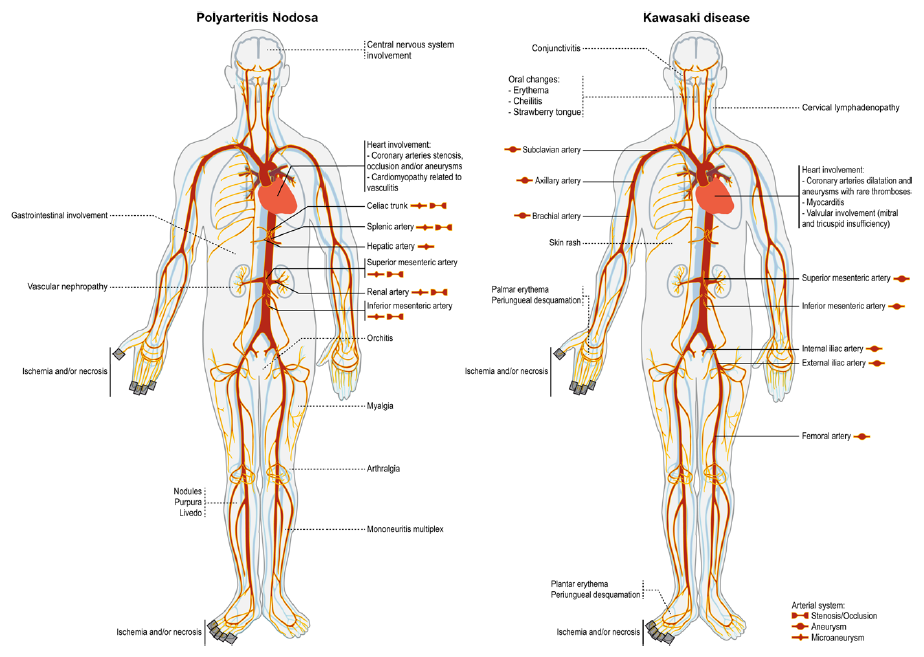
Figure 2. Representative pattern of predominant vascular and organ lesions in medium vessel vasculitis (Doi: 10.1161/CIRCULATIONAHA.120.046657)
3. Small Vessel Vasculitis (SVV)
Involves small arteries, capillaries, and venules. This is the most common clinical subtype of vasculitis and is often associated with abnormal autoantibody levels. Common types include:
- ANCA-Associated Vasculitis (AAV): A core subtype encompassing Granulomatosis with Polyangiitis (GPA), Microscopic Polyangiitis (MPA), and Eosinophilic Granulomatosis with Polyangiitis (EGPA). These are typified by respiratory tract involvement, renal involvement, and asthma with eosinophilia, respectively.
- IgA Vasculitis (Henoch-Schönlein Purpura): Most frequent in children and adolescents, classically presenting with symmetric purpura on the lower extremities, arthralgia, and gastrointestinal symptoms. Some cases may involve the kidneys, causing immune complex-mediated renal injury.
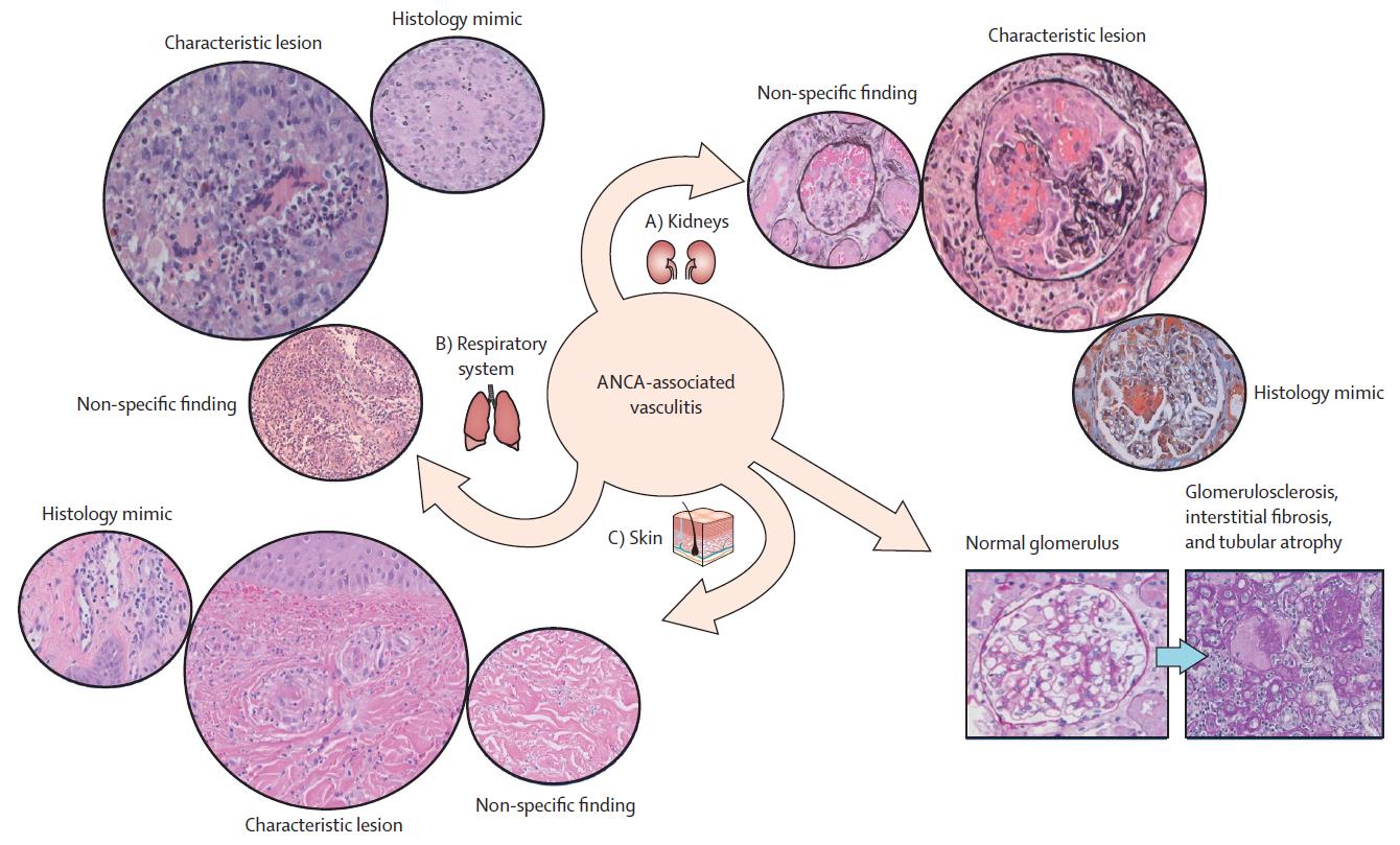
Figure 3. The effect of ANCA-associated vasculitis (Doi: 10.1016/S0140-6736(23)01736-1)
III. Decoding Pathogenesis: Immune-Inflammatory Cascades and Key Signaling Pathways
The core pathogenesis of vasculitis involves an inflammatory cascade initiated by abnormal immune system activation. Multiple interconnected signaling pathways collaboratively mediate blood vessel wall damage. The key mechanisms and core pathways are as follows:
1. Core Pathogenic Mechanisms
Immune system dysregulation is central to vasculitis development. On the innate immunity level, aberrantly activated neutrophils release Neutrophil Extracellular Traps (NETs). While NETs are a defensive mechanism for pathogen clearance, their excessive formation or impaired clearance can damage vascular endothelial cells, a key pathological step in AAV. On the adaptive immunity level, abnormal activation of T cells and B cells leads to the overproduction of autoantibodies (e.g., ANCA) and pro-inflammatory cytokines, sustaining a persistent immune-inflammatory response that targets and damages vessel walls.
Recent research indicates that in vasculitis, vascular endothelial cells are not merely passive targets of inflammatory injury but actively participate in immune response regulation. Under inflammatory stimulation, endothelial cells upregulate adhesion molecules and chemotactic signals, promoting the recruitment and retention of neutrophils and T cells within the vessel wall. Within a sustained inflammatory microenvironment, their immunophenotype alters, potentially further amplifying local inflammation. This endothelial cell-mediated immunomodulatory effect is believed to be closely linked to the maintenance of chronic vascular wall inflammation and vascular remodeling, holding particular significance in the progression of large vessel vasculitis.
At the adaptive immunity level, T cell abnormalities represent a broader functional imbalance rather than a singular activation state. A persistently enhanced pro-inflammatory T cell response sustains the vascular wall inflammatory microenvironment and promotes long-term inflammatory cell infiltration. Concurrently, impaired immunoregulatory functions hinder the effective termination of the inflammatory response. In AAV, aberrant interactions between T cells and B cells are thought to be closely associated with the continuous production of autoantibodies and maintenance of disease activity, collectively driving chronicity and relapse.
2. Key Signaling Pathways
- NF-κB Pathway: A central regulatory pathway for inflammatory responses. It can be activated by stimuli such as infectious agents and autoantibodies, initiating the transcription of numerous downstream pro-inflammatory genes and the synthesis of cytokines like TNF-α and IL-6, exacerbating inflammatory cell infiltration and tissue damage in the vessel wall.
- MAPK Pathway: Includes major branches such as p38, JNK, and ERK. It is responsible for transmitting inflammatory signals and regulating immune cell activation, proliferation, and cytokine secretion. It plays a crucial role in neutrophil activation and NETs formation, serving as a key pathway for amplifying inflammatory responses.
- JAK-STAT Pathway: The core pathway mediating cytokine signal transduction. Upon binding of pro-inflammatory cytokines like IL-6 and interferons to their cell surface receptors, JAK kinases are activated, leading to the phosphorylation of STAT proteins and their translocation into the nucleus to initiate gene expression related to immune cell proliferation and inflammatory factor release. This pathway is aberrantly activated in large vessel vasculitis and represents an important potential target for therapy.
- Complement Pathway: A vital component of the innate immune system. Activation leads to the generation of C5a, which recruits neutrophils to sites of vascular injury via chemotaxis, aggravating local inflammation. It plays a key mediating role in renal injury in AAV.
- mTOR Pathway: A central pathway regulating immune cell metabolism, proliferation, and function. Its abnormal activation in large vessel vasculitis can lead to excessive T cell activation and infiltration into the vessel wall, promoting granuloma formation and vascular remodeling, thereby worsening pathological changes like vascular stenosis.
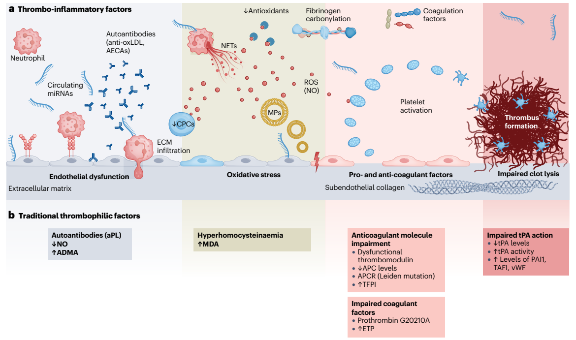
Figure 4. The main pathogenetic mechanisms in vasculitis (Doi: 10.1038/s41584-022-00880-7)
IV. Mechanisms and Targets: Advances in Precision Therapy for Vasculitis
With deeper understanding of vasculitis pathogenesis, the treatment paradigm is shifting from non-specific therapies like glucocorticoids combined with conventional immunosuppressants towards precision-targeted therapies. Current research and clinical focus areas include the following key targets:
1. Cytokine Targets
- TNF-α: A pivotal pro-inflammatory cytokine mediating inflammatory cell infiltration and vascular endothelial damage. TNF-α inhibitors (e.g., Infliximab, Adalimumab) have shown definitive anti-inflammatory efficacy in refractory cases like Takayasu arteritis and polyarteritis nodosa.
- IL-6: A key cytokine mediating systemic inflammation and immune cell activation. The anti-IL-6 receptor monoclonal antibody, Tocilizumab, has achieved breakthrough progress in treating Giant Cell Arteritis, significantly reducing glucocorticoid doses and improving relapse-free survival rates. It has become an important clinical treatment option.
- IL-5: A cytokine specifically regulating eosinophil activation, proliferation, and survival. Mepolizumab, an anti-IL-5 monoclonal antibody, is the first targeted drug approved for Eosinophilic Granulomatosis with Polyangiitis (EGPA). It precisely inhibits eosinophil-mediated inflammation, improving clinical symptoms.
2. Immune Cell Targets
- CD20: A B-cell surface-specific marker. The anti-CD20 monoclonal antibody Rituximab specifically depletes B cells, reducing the production of autoantibodies like ANCA. Its efficacy is comparable to traditional cyclophosphamide with a better safety profile, establishing it as a first-line targeted therapy for AAV.
- Neutrophils/NETs: Targeted interventions against NETs formation or clearance mechanisms represent a novel research direction for AAV. Agents like Nrf2 activators and CypD inhibitors may mitigate vascular endothelial damage by inhibiting NETs formation or promoting their clearance, currently in preclinical or early clinical research stages.
3. Signaling Pathway Targets
- C5a Receptor: A key regulatory target in the complement pathway. The small molecule inhibitor Avacopan specifically blocks the binding of C5a to its receptor on neutrophils, inhibiting neutrophil recruitment and activation. It has shown promising efficacy in treating AAV and can reduce glucocorticoid dependence, having received clinical approval.
- JAK: The core kinase of the JAK-STAT pathway. JAK inhibitors like Tofacitinib and Baricitinib broadly inhibit multiple cytokine signaling pathways, suppressing T cell activation and vascular wall inflammatory infiltration. They hold potential therapeutic value for refractory large vessel vasculitis and AAV, with related clinical trials underway.
- mTOR: The mTOR pathway inhibitor Sirolimus (Rapamycin) inhibits excessive immune cell proliferation and modulates vascular wall inflammation and remodeling processes. It shows potential application prospects in intervening vascular pathologies of large vessel vasculitis, offering a new strategy for addressing pathological changes like vascular stenosis.
V. Current Status and Future Directions in Vasculitis Diagnosis and Treatment
Vasculitis represents a group of immune-mediated diseases marked by strong heterogeneity and complex pathogenesis. Its core pathological feature is immune-inflammatory infiltration and damage of the blood vessel wall, with clinical manifestations varying significantly based on the size and location of affected vessels. The classification system based on the Chapel Hill Consensus provides a unified standard for clinical diagnosis and research. The inflammatory cascade triggered by immune system imbalance and the aberrant regulation of signaling pathways like NF-κB, JAK-STAT, and complement are key to unraveling the disease's nature. In recent years, significant progress has been made in precision therapies targeting cytokines, immune cells, and signaling pathways, with several targeted drugs approved for clinical use, greatly improving patient prognosis. Looking forward, further exploration of disease molecular mechanisms, discovery of novel therapeutic targets, optimization of multi-target combination strategies, and refinement of individualized treatment will form the core directions of vasculitis research. These efforts hold promise for providing more effective and safer solutions for clinical management while offering new avenues to address challenges like disease relapse and intervention in refractory cases.
abinScience Vasculitis Research Solutions
1. Antibodies
| Product Name |
Catalog No. |
| InVivoMAb Anti-Human TNFa/TNF-alpha (Iv0050) |
HF879010 |
| Anti-IL17A Polyclonal Antibody |
HS856014 |
| InVivoMAb Anti-Human IL5 (Iv0021) |
HY218010 |
| Anti-JAK1 Polyclonal Antibody |
HB829014 |
| Anti-Human CD20/MS4A1 Antibody (SAA2511) |
HY257033 |
| Research Grade Anti-Human CD88/C5AR1 Antibody (G2_anti-C5aR) |
HB064056 |
| Anti-Human CD126/IL6R Antibody (SAA0048) |
HY583107 |
| Anti-Human IL12A/IL-12 p35 Antibody (SAA0380) |
HB936107 |
View more Vasculitis Antibodies
2. Assay Kits
View more Vasculitis Assay Kits
3. Research Biosimilars
| Target |
Product Name |
Catalog No. |
| CD20 |
Research Grade Ofatumumab |
HY257466 |
| Research Grade Rituximab |
HY257446 |
| CD88/C5AR1 |
Research Grade Avdoralimab |
HB064016 |
| Research Grade Izastobart |
HB064046 |
| IL-6R |
Research Grade Tocilizumab |
HY583026 |
| IL-5 |
Research Grade Mepolizumab |
HY218026 |
| TNFa |
Research Grade Etanercept |
HF879296 |
| Research Grade Adalimumab |
HF879026 |
| IL17A |
Research Grade Betinukibart |
HS856236 |
| Research Grade Secukinumab |
HS856026 |
View more Vasculitis Research Biosimilars
abinScience offers high-quality research tools covering core mechanisms and targets in vasculitis research. This includes specific antibodies against key molecules like TNF-α, IL-6, and CD20, as well as ADA (Anti-Drug Antibody) and PK (Pharmacokinetic) assay kits. These tools precisely support mechanistic studies in core areas such as interferon signaling pathways, abnormal B-cell activation, complement activation, and NETs formation. They provide reliable data support for immunogenicity assessment and pharmacokinetic analysis of vasculitis-targeted drugs, aiding in accelerating drug development and translational applications.
Need help selecting reagents for your vasculitis project? Contact us: [email protected] | +86-27-65523339
References
- Cannon L, Wu EY. Recent Advances in Pediatric Vasculitis. Rheum Dis Clin North Am. 2021;47(4):781-796. doi:10.1016/j.rdc.2021.07.007
- Teague HL, Ahlman MA, Alavi A, et al. Unraveling Vascular Inflammation: From Immunology to Imaging. J Am Coll Cardiol. 2017;70(11):1403-1412. doi:10.1016/j.jacc.2017.07.750
- Saadoun D, Vautier M, Cacoub P. Medium- and Large-Vessel Vasculitis. Circulation. 2021;143(3):267-282. doi:10.1161/CIRCULATIONAHA.120.046657
- Hoffman GS, Calabrese LH. Vasculitis: determinants of disease patterns. Nat Rev Rheumatol. 2014;10(8):454-462. doi:10.1038/nrrheum.2014.89
- Sunderkötter CH, Zelger B, Chen KR, et al. Nomenclature of Cutaneous Vasculitis. Arthritis Rheumatol. 2018;70(2):171-184. doi:10.1002/art.40375
- Kronbichler A, Bajema IM, Bruchfeld A, et al. Diagnosis and management of ANCA-associated vasculitis. Lancet. 2024;403(10427):683-698. doi:10.1016/S0140-6736(23)01736-1
- Bettiol A, Alibaz-Oner F, Direskeneli H, et al. Vascular Behçet syndrome: from pathogenesis to treatment. Nat Rev Rheumatol. 2023;19(2):111-126. doi:10.1038/s41584-022-00880-7
For Research Use Only. Not for use in diagnostic or therapeutic procedures.


 中文
中文 English
 한국어
한국어 日本語
日本語 Español
 Français
 Русский


