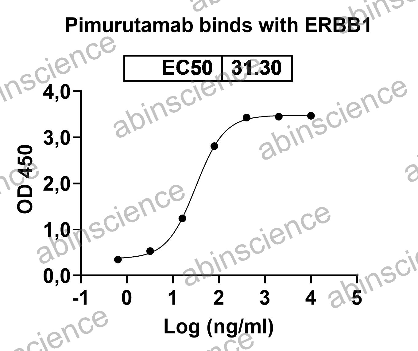
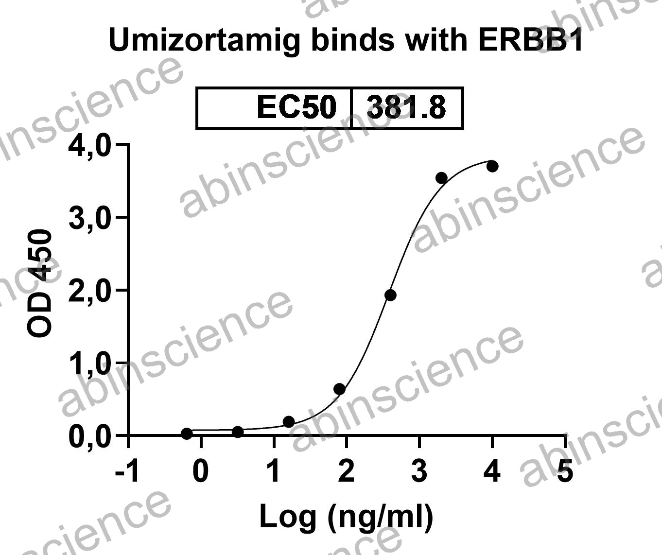
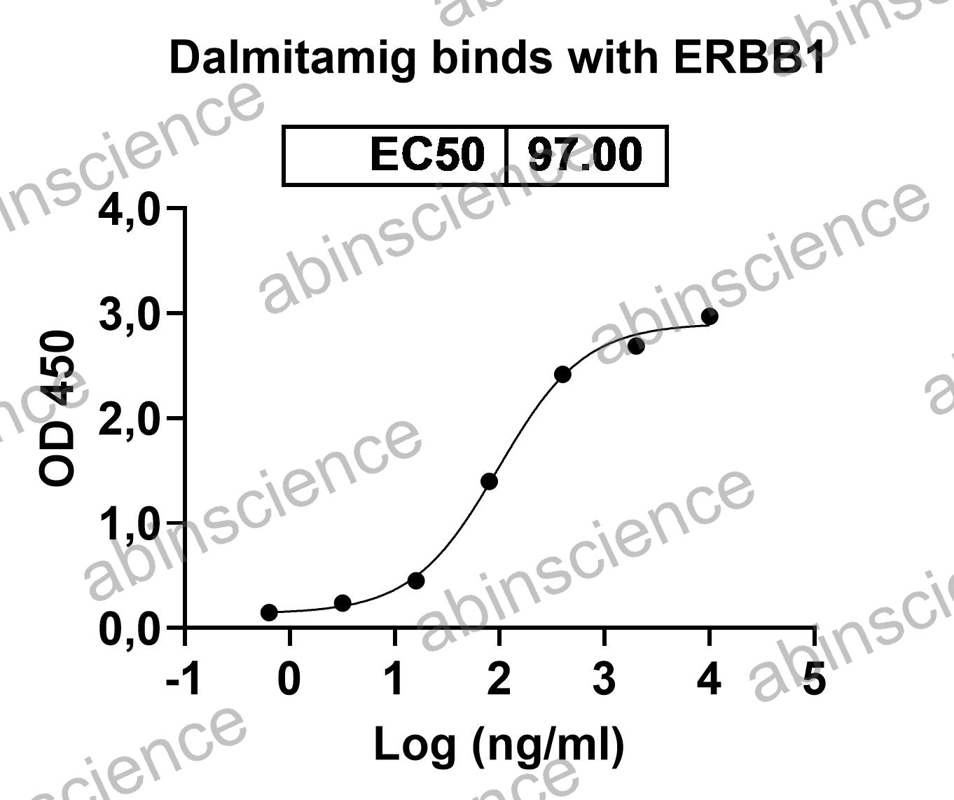
EGFR (Epidermal Growth Factor Receptor / ErbB1 / HER1) is the founding member of the ErbB receptor tyrosine kinase family and one of the most extensively validated oncology targets. EGFR-activating mutations and amplification drive tumor growth in non-small cell lung cancer (NSCLC), colorectal cancer (CRC), head and neck squamous cell carcinoma (HNSCC), and glioblastoma. Approved EGFR-targeting therapies span multiple modalities: therapeutic antibodies Cetuximab (Erbitux) and Panitumumab (Vectibix), tyrosine kinase inhibitors (Osimertinib, Erlotinib, Gefitinib), and the bispecific antibody Amivantamab (EGFR × MET). abinScience offers recombinant EGFR proteins, anti-EGFR antibodies, EGF ligand, biosimilar reference standards, and ELISA kits.
EGFR Biology and Oncogenic Signaling
EGFR is a ~170 kDa single-pass transmembrane receptor with an extracellular domain (ECD) comprising four subdomains (I-IV), a transmembrane helix, and an intracellular tyrosine kinase domain. In the inactive state, EGFR adopts a tethered conformation where subdomain II (dimerization arm) is masked by intramolecular contact with subdomain IV. Ligand binding (EGF, TGF-α, amphiregulin) to subdomains I and III releases the tether, exposing the dimerization arm for homo- or heterodimerization with HER2, HER3, or HER4, triggering kinase activation and downstream RAS/MAPK, PI3K/AKT, and JAK/STAT3 signaling.
EGFR-driven cancers arise through distinct mechanisms depending on tumor type. In NSCLC, activating kinase domain mutations (exon 19 deletion, L858R, exon 20 insertions) render the receptor constitutively active independent of ligand. In CRC and HNSCC, EGFR is typically wild-type but overexpressed or amplified, and ligand-dependent signaling drives proliferation. This distinction determines the therapeutic approach: TKIs for mutant EGFR (NSCLC), therapeutic antibodies for wild-type EGFR overexpression (CRC, HNSCC). The third-generation TKI Osimertinib was specifically designed to overcome the T790M gatekeeper resistance mutation that emerged with first-generation TKIs.
Key Research Applications
Anti-EGFR Antibody & Bispecific Development Recombinant EGFR ECD proteins (wild-type and mutant variants) for screening anti-EGFR therapeutic antibodies by SPR/BLI. EGFR × MET bispecific format studies using matched EGFR and MET proteins.
Cetuximab Biosimilar Programs Research-grade Cetuximab and Panitumumab biosimilars for analytical comparability, ADCC potency bioassays, ligand-blocking ELISA, and glycan profiling. Cetuximab biosimilar development requires careful α-Gal epitope characterization.
EGFR Signaling & Pathway Studies Bioactive EGF, TGF-α, and amphiregulin proteins for stimulating EGFR phosphorylation and downstream signaling. Anti-phospho-EGFR antibodies (pY1068, pY1173) for pathway activation monitoring by WB and ELISA.
IHC, FISH & Companion Diagnostics Anti-EGFR antibodies validated for IHC on FFPE tissue. EGFR IHC and FISH are used for patient selection in CRC (KRAS wild-type) and NSCLC (mutation testing). Anti-EGFR vIII antibodies for glioblastoma-specific variant detection.
EGFR Product Selection Guide
| Application |
Recommended Product |
Format |
| SPR/BLI binding kinetics |
Recombinant EGFR ECD protein |
Avi-tagged or biotinylated |
| EGF/EGFR ligand-blocking ELISA |
EGFR-Fc + EGF protein |
Fc-chimera + His-tagged |
| ADCC potency assay |
Cetuximab biosimilar + EGFR⁺ target cells |
Research-grade RUO |
| EGFR signaling / proliferation |
Bioactive EGF or TGF-α protein |
Carrier-free, low endotoxin |
| IHC (FFPE tissue) |
Anti-EGFR monoclonal antibody |
Unconjugated, IHC-validated |
| EGFRvIII detection (glioblastoma) |
Anti-EGFRvIII specific antibody |
Unconjugated |
| Flow cytometry |
Anti-EGFR antibody |
PE or APC conjugated |
| Serum sEGFR quantification |
EGFR ELISA kit |
Sandwich ELISA |
| PK/ADA ELISA |
Anti-idiotype pair + EGFR calibrator |
Capture/detection pair |
| Analytical comparability |
Cetuximab or Panitumumab biosimilar |
Research-grade RUO |
EGFR FAQs
What is the difference between Cetuximab and Panitumumab?
Cetuximab is a chimeric (mouse/human) IgG1 antibody that binds EGFR subdomain III, blocking ligand binding and enabling Fc-mediated ADCC. It is produced in murine SP2/0 cells and carries α-1,3-galactose (α-Gal) glycan epitopes, which can cause hypersensitivity reactions in patients with pre-existing anti-α-Gal IgE. Panitumumab is a fully human IgG2 antibody that also binds subdomain III but lacks ADCC activity (IgG2 Fc) and is produced in CHO cells without α-Gal. Both require KRAS/NRAS wild-type status in CRC patients — downstream RAS mutations render EGFR blockade ineffective.
What is the EGFR mutation landscape in NSCLC?
The most common activating EGFR mutations in NSCLC are exon 19 deletions (~45%) and L858R point mutation in exon 21 (~40%) — both are sensitive to TKI therapy. Exon 20 insertions (~10%) are generally TKI-resistant but targetable by Amivantamab (bispecific antibody). The T790M gatekeeper mutation (exon 20) is the dominant resistance mechanism to 1st/2nd-generation TKIs, overcome by Osimertinib. C797S is an emerging resistance mechanism to Osimertinib. For research, we offer wild-type and mutant EGFR ECD proteins, and anti-EGFR antibodies suitable for detecting both wild-type and mutant receptor by WB and IHC.
What is EGFRvIII and why is it important?
EGFRvIII (variant III) is a deletion mutant lacking exons 2-7 (267 amino acids from the extracellular domain), creating a constitutively active receptor that cannot bind ligand. EGFRvIII is found in ~30% of glioblastomas and a subset of HNSCC. Its tumor-specific expression (absent on normal tissues) makes it an attractive target for CAR-T therapy, ADCs, and tumor-specific vaccines (Rindopepimut). Anti-EGFRvIII antibodies that recognize the unique junction epitope do not cross-react with wild-type EGFR.
Why is α-Gal glycosylation important for Cetuximab biosimilar development?
Cetuximab is produced in murine SP2/0 cells that add α-1,3-galactose (α-Gal) residues to the Fc glycans — a non-human glycan structure. Patients with pre-existing anti-α-Gal IgE antibodies (common in certain geographic regions due to tick bites/α-Gal syndrome) can develop severe anaphylaxis. Cetuximab biosimilar development must carefully characterize α-Gal content and demonstrate comparable levels to the reference product. Some biosimilar manufacturers produce Cetuximab in CHO cells (which lack the α-Gal transferase), but this creates a glycan profile different from the reference, complicating regulatory comparability.
Where do Cetuximab and Panitumumab bind on EGFR?
Both Cetuximab and Panitumumab bind EGFR subdomain III (the ligand-binding domain), with partially overlapping but non-identical epitopes. They directly compete with EGF and TGF-α for receptor binding, preventing ligand-induced dimerization and signaling. This is distinct from HER2-targeting antibodies: Trastuzumab binds subdomain IV and Pertuzumab binds subdomain II. For epitope mapping of novel anti-EGFR candidates, individual EGFR subdomain proteins and competitive binding assays with Cetuximab/Panitumumab can determine epitope overlap.
Key References
1. Cunningham D, et al. (2004) Cetuximab monotherapy and cetuximab plus irinotecan in irinotecan-refractory metastatic colorectal cancer. N Engl J Med. 351(4):337-345. PMID: 15269313
2. Soria JC, et al. (2018) Osimertinib in untreated EGFR-mutated advanced non-small-cell lung cancer. N Engl J Med. 378(2):113-125. PMID: 29151359
3. Li S, et al. (2005) Structural basis for inhibition of the epidermal growth factor receptor by cetuximab. Cancer Cell. 7(4):301-311. PMID: 15837620


 中文
中文 English
 한국어
한국어 日本語
日本語 Español
 Français
 Русский