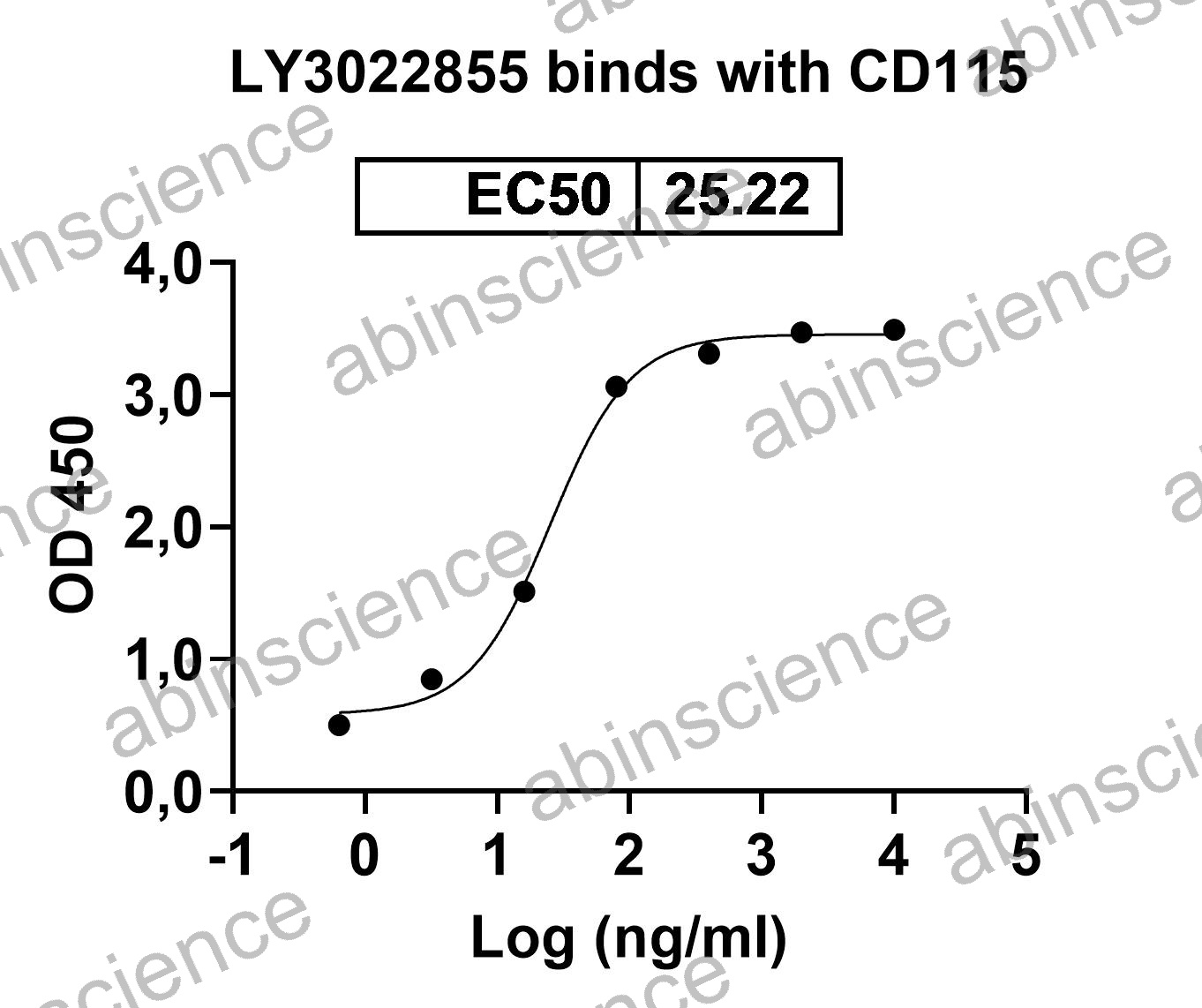
CSF1R (Colony Stimulating Factor 1 Receptor / CD115 / M-CSFR) is a receptor tyrosine kinase that drives the differentiation, survival, and function of monocytes, macrophages, osteoclasts, and microglia. CSF1R is activated by two ligands — CSF1 (M-CSF) and IL-34 — and is a key therapeutic target for reprogramming tumor-associated macrophages (TAMs) in immuno-oncology. The CSF1R inhibitor Pexidartinib (Turalio) is FDA-approved for tenosynovial giant cell tumor (TGCT), and multiple anti-CSF1R antibodies are in clinical trials for solid tumors. abinScience offers recombinant CSF1R and CSF1 proteins, anti-CSF1R antibodies, and ELISA kits.
CSF1R Biology and the TAM Axis
CSF1R is a ~150 kDa type III receptor tyrosine kinase with five extracellular Ig-like domains. It is expressed on monocytes, macrophages, dendritic cell precursors, osteoclasts, and microglia. CSF1R has two ligands: CSF1 (M-CSF), which is widely expressed and drives steady-state macrophage homeostasis, and IL-34, which has more restricted expression (skin, brain) and preferentially maintains Langerhans cells and microglia.
In the tumor microenvironment, tumor-derived CSF1 recruits and programs macrophages toward an immunosuppressive M2-like (TAM) phenotype that promotes angiogenesis, matrix remodeling, and T-cell exclusion. Blocking CSF1R — with either small-molecule kinase inhibitors (Pexidartinib, PLX3397) or anti-CSF1R antibodies (Cabiralizumab, Emactuzumab) — can deplete TAMs or reprogram them toward a pro-inflammatory M1 phenotype, making CSF1R blockade synergistic with PD-1/PD-L1 checkpoint inhibitors.
Key Research Applications
Anti-CSF1R Drug Development Recombinant CSF1R ECD proteins for screening anti-CSF1R therapeutic antibodies by SPR/BLI and competitive ELISA. CSF1 and IL-34 ligand proteins for receptor-ligand blocking assays.
Macrophage Differentiation & Polarization Bioactive CSF1 (M-CSF) protein for differentiating monocytes into macrophages (7-day M-CSF protocol). Anti-CSF1R blocking antibodies for studying TAM reprogramming from M2 to M1 phenotype in co-culture tumor models.
Microglia & Neuroinflammation CSF1 and IL-34 proteins for microglial maintenance and activation studies. Anti-CSF1R antibodies for microglial depletion models in neurodegeneration research (Alzheimer's, ALS, glioblastoma).
Bone Biology & Osteoclast Research CSF1 + RANKL proteins for osteoclast differentiation from bone marrow precursors. Anti-CSF1R antibodies for studying osteoclast-mediated bone resorption in osteoporosis and bone metastasis models.
CSF1R Product Selection Guide
| Application |
Recommended Product |
Format |
| SPR/BLI binding kinetics |
Recombinant CSF1R ECD protein |
Avi-tagged or biotinylated |
| CSF1/CSF1R blocking ELISA |
CSF1R-Fc + CSF1 protein |
Fc-chimera + His-tagged |
| Monocyte → macrophage differentiation |
Bioactive CSF1 (M-CSF) |
Carrier-free, low endotoxin |
| Osteoclast differentiation |
CSF1 + RANKL proteins |
Carrier-free, bioassay-validated |
| Flow cytometry / monocyte profiling |
Anti-CD115/CSF1R antibody |
PE or APC conjugated |
| IL-34/CSF1R selectivity testing |
CSF1R + CSF1 + IL-34 proteins |
His-tagged (matched format) |
| PK/ADA ELISA |
Anti-CSF1R antibody pair + calibrator |
Capture/detection pair |
CSF1R FAQs
What is the difference between CSF1 (M-CSF) and IL-34 as CSF1R ligands?
CSF1 (M-CSF) is ubiquitously expressed and is the primary ligand for monocyte/macrophage homeostasis. IL-34 has restricted expression — mainly in the skin (keratinocytes) and brain (neurons) — and preferentially maintains Langerhans cells and microglia. Both bind CSF1R but at non-overlapping sites, and they share no sequence homology. Anti-CSF1R antibodies block both ligands, while anti-CSF1 antibodies block only CSF1, leaving IL-34-dependent populations (microglia, Langerhans cells) intact. This distinction has safety implications for therapeutic strategies.
Does CSF1R blockade deplete or reprogram macrophages?
Both, depending on the approach and context. Complete CSF1R blockade (high-dose small-molecule inhibitor or depleting anti-CSF1R antibody) eliminates macrophages by removing their survival signal. Partial or transient blockade can reprogram TAMs from an immunosuppressive M2 phenotype toward a pro-inflammatory M1 phenotype without full depletion. In preclinical tumor models, combination with anti-PD-1 works better with reprogramming than depletion, as residual pro-inflammatory macrophages can support anti-tumor immunity.
How do I differentiate monocytes into macrophages using M-CSF?
Isolate CD14⁺ monocytes from PBMCs by magnetic bead selection. Culture in RPMI + 10% FBS supplemented with 25-50 ng/mL recombinant M-CSF (CSF1) for 7 days, refreshing medium and cytokine on day 3-4. This produces M0 (unpolarized) macrophages. For M1 polarization, add IFN-γ + LPS on day 6. For M2 polarization, add IL-4 + IL-13. Verify differentiation by flow cytometry: CD14⁺CD68⁺CD163⁻ (M1) or CD14⁺CD68⁺CD163⁺CD206⁺ (M2).
What is the role of CSF1R in microglia and neurodegeneration?
Microglia are the brain's resident macrophages and depend on CSF1R signaling (primarily via IL-34 in the CNS) for survival. CSF1R inhibitor-mediated microglial depletion (using PLX5622 or BLZ945 in preclinical models) is a widely used tool for studying microglial contributions to Alzheimer's disease, ALS, traumatic brain injury, and glioblastoma. Upon inhibitor withdrawal, microglia rapidly repopulate from surviving progenitors. In glioblastoma, CSF1R blockade reprograms tumor-associated microglia/macrophages and synergizes with anti-PD-1 therapy.
How do I differentiate osteoclasts from bone marrow precursors?
Culture mouse bone marrow cells or human CD14⁺ monocytes with M-CSF (25 ng/mL) + RANKL (50-100 ng/mL) for 5-7 days. M-CSF provides survival signaling through CSF1R, while RANKL drives osteoclast commitment and fusion. Mature osteoclasts are identified as large multinucleated (3+ nuclei) TRAP-positive cells. Functional resorption can be assessed on dentine discs or calcium phosphate-coated plates. Anti-CSF1R blocking antibodies should inhibit both M-CSF-dependent survival and osteoclast formation.
Key References
1. Cannarile MA, et al. (2017) Colony-stimulating factor 1 receptor (CSF1R) inhibitors in cancer therapy. J Immunother Cancer. 5(1):53. PMID: 28716061
2. Elmore MR, et al. (2014) Colony-stimulating factor 1 receptor signaling is necessary for microglia viability. J Neurosci. 34(33):11929-11947. PMID: 25122893
3. Lin H, et al. (2008) Discovery of a cytokine and its receptor by functional screening of the extracellular proteome. Science. 320(5877):807-811. PMID: 18467591


 中文
中文 English
 한국어
한국어 日本語
日本語 Español
 Français
 Русский