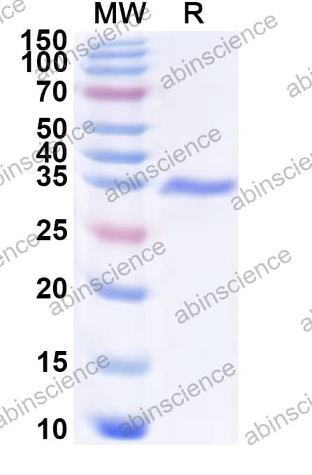
Anti-HHAV Protease 3C Polyclonal Antibody (VK802014) is a rabbit polyclonal antibody detecting Protease 3C. Suitable for Simian hepatitis A virus genotype V, Human hepatitis A virus genotype IIA, Human hepatitis A virus genotype IB, Human hepatitis A virus genotype IIIB, Human hepatitis A virus genotype IIB, Human hepatitis A virus genotype IA, and Human hepatitis A virus genotype IIIA.
Highlights
●Multi-Species — Cross-reactive for translational research.
Species reactivity
Hepatitis A virus (HAV)
Applications
ELISA, IHC, WB
Host species
Rabbit
Isotype
IgG
Clonality
Polyclonal
Immunogen
E. coli - derived recombinant HHAV Protease 3C (Ser1520-Gln1738).
Target
Assembly signal 2A, Capsid protein VP0, Capsid protein VP1, Capsid protein VP2, Capsid protein VP3, Capsid protein VP4, EC:2.7.7.48, EC:3.4.22.28, EC:3.6.1.15, Genome polyprotein, P1A, P1B, P1C, P1D, P2B, P2C, P3, P3A, P3B, P3C, P3D-POL, Picornain 3C, Protease 3C, Protein 2B, Protein 2BC, Protein 2C, Protein 3A, Protein 3AB, Protein 3ABC, Protein 3ABCD, Protein 3B, Protein 3CD, Protein VP1-2A, RNA-directed RNA polymerase 3D-POL, VP4-VP2, VPX, VPg, Viral protein genome-linked, Virion protein 1, Virion protein 2, Virion protein 3, Virion protein 4, pX
Please refer to the specific buffer information in the hardcopy of datasheet or the lot-specific COA.
Product Usage Information
Application
Dilution
ELISA
1:5000-1:20000
IHC
1:50-1:500
WB
1:500-1:2000
Background
Genome polyprotein (Protease 3C) is a ~251 kDa protein. Capsid proteins VP1, VP2, and VP3 form a closed capsid enclosing the viral positive strand RNA genome. All these proteins contain a beta-sheet structure called beta-barrel jelly roll. Together they form an icosahedral capsid (T=3) composed of 60 copies of each VP1, VP2, and VP3, with a diameter of approximately 300 Angstroms. VP1 is situated at the 12 fivefold axes, whereas VP2 and VP3 are located at the quasi-sixfold axes. The naked capsid interacts with the host receptor HAVCR1 to provide virion attachment to and probably entry into the target cell.

 中文
中文 English
 한국어
한국어 日本語
日本語 Español
 Français
 Русский