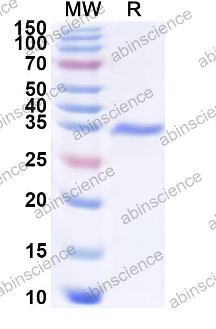
ELISA, Immunogen, SDS-PAGE, WB, Bioactivity testing in progress
Species
Hepatitis A virus (HAV)
Nature
Recombinant
Endotoxin level
Please contact with the lab for this information.
Purity
>90% as determined by SDS-PAGE.
Form
Lyophilized
Storage buffer
Lyophilized from a solution in PBS pH 7.4, 1 mM EDTA, 4% Trehalose, 1% Mannitol.
Please refer to the specific buffer information in the hardcopy of datasheet or the lot-specific COA.
Reconstitution
Reconstitute in sterile water for a stock solution. A copy of datasheet will be provided with the products, please refer to it for details.
Shipping
In general, proteins are provided as lyophilized powder/frozen liquid. They are shipped out with dry ice/blue ice unless customers require otherwise.
Stability and Storage
Use a manual defrost freezer and avoid repeated freeze thaw cycles. Store at 2 to 8°C for frequent use. Store at -20 to -80°C for twelve months from the date of receipt.
Alternate Names
Assembly signal 2A, Capsid protein VP0, Capsid protein VP1, Capsid protein VP2, Capsid protein VP3, Capsid protein VP4, EC:2.7.7.48, EC:3.4.22.28, EC:3.6.1.15, Genome polyprotein, P1A, P1B, P1C, P1D, P2B, P2C, P3, P3A, P3B, P3C, P3D-POL, Picornain 3C, Protease 3C, Protein 2B, Protein 2BC, Protein 2C, Protein 3A, Protein 3AB, Protein 3ABC, Protein 3ABCD, Protein 3B, Protein 3CD, Protein VP1-2A, RNA-directed RNA polymerase 3D-POL, VP4-VP2, VPX, VPg, Viral protein genome-linked, Virion protein 1, Virion protein 2, Virion protein 3, Virion protein 4, pX
Background
Genome polyprotein is a ~251 kDa protein. Capsid proteins VP1, VP2, and VP3 form a closed capsid enclosing the viral positive strand RNA genome. All these proteins contain a beta-sheet structure called beta-barrel jelly roll. Together they form an icosahedral capsid (T=3) composed of 60 copies of each VP1, VP2, and VP3, with a diameter of approximately 300 Angstroms. VP1 is situated at the 12 fivefold axes, whereas VP2 and VP3 are located at the quasi-sixfold axes. The naked capsid interacts with the host receptor HAVCR1 to provide virion attachment to and probably entry into the target cell.

 中文
中文 English
 한국어
한국어 日本語
日本語 Español
 Français
 Русский