In early October, pig farms in Japan reported a series of classical swine fever (CSF; known in Japan as "ton-netsu") outbreaks. On October 7, Japan's Ministry of Agriculture, Forestry and Fisheries confirmed CSF at a pig farm in Ozu, Ehime Prefecture, marking the country's 106th case; the farm held about 4,000 pigs. Just three days later, infection was confirmed at a farm in Shibukawa, Gunma Prefecture, the 107th case, involving the affected farm and one linked farm with roughly 2,800 pigs in total. Meanwhile, new infections in wild boar continue to be detected: Ibaraki Prefecture was still reporting positive wild boar in October and has deployed oral vaccine baits in 22 municipalities. Since CSF re-emerged in Gifu Prefecture in 2018, CSF-positive wild boar have been found in 43 prefectures across Japan, and outbreaks on farms have continued to recur.
The strain responsible for Japan's epidemic since 2018 belongs to subgenotype 2.1d, which was first isolated in China in 2014–2015 and typically causes chronic or moderate infections. Moderately virulent strains produce atypical clinical signs and a prolonged disease course, and infected animals can shed virus for long periods, which is one reason the virus has persisted in wild boar populations. Other studies suggest that the C-strain used in bait vaccines may protect less effectively against 2.1d sublineage strains than against other 2.1 subgenotypes. Understanding the molecular features of this virus is the foundation for developing diagnostics and vaccines.
01Virus Classification and Genome Organization
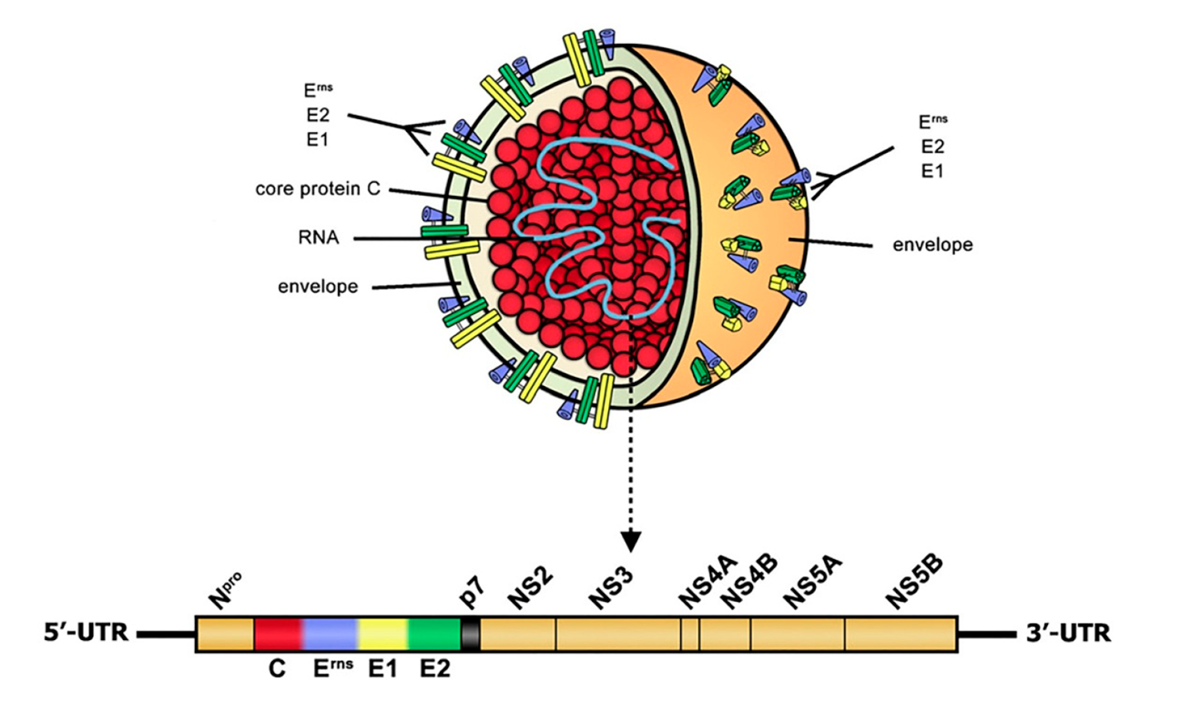
Classical swine fever virus (CSFV) belongs to the genus Pestivirus in the family Flaviviridae, together with bovine viral diarrhea virus (BVDV) and border disease virus (BDV). Its only natural hosts are domestic pigs and wild boar, and it does not infect humans. Pestiviruses are small enveloped viruses with a single-stranded, positive-sense RNA genome that lacks a 5' cap and a 3' poly(A) tail. The CSFV genome is approximately 12.5 kb and contains a single open reading frame encoding a polyprotein of about 3,900 amino acids. Co- and post-translational processing by host and viral proteases yields 11 to 12 mature proteins, arranged in the order Npro-C-Erns-E1-E2-p7-NS2-NS3-NS4A-NS4B-NS5A-NS5B. C, Erns, E1 and E2 are structural proteins, and the rest are non-structural proteins. The 5' untranslated region contains an internal ribosome entry site (IRES) that mediates cap-independent translation initiation.
02Structural Proteins
E2: The Major Immunogen and a Key Entry Protein
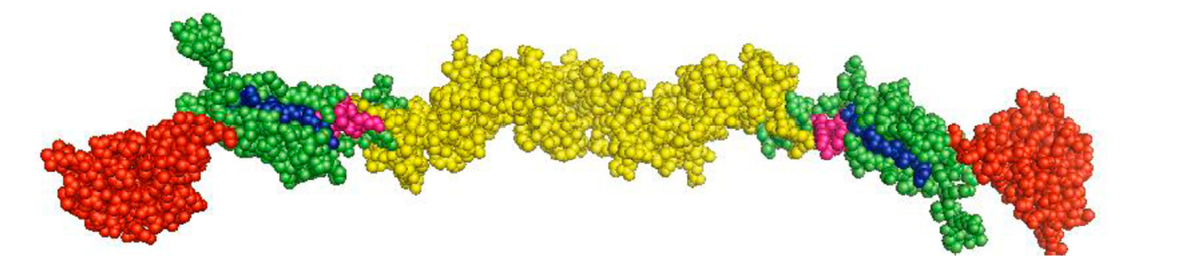
E2 is the most immunogenic CSFV protein. It induces protective neutralizing antibodies and, on its own, can confer protection in animals. Viral mutants with partial or complete deletion of E2 are not viable, indicating that E2 is essential to the viral life cycle; it is also an important virulence determinant. On the virion, E2 forms disulfide-linked homodimers of about 100 kDa and can also form heterodimers with E1.
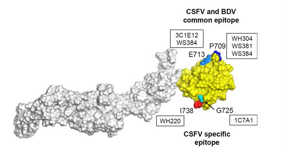
The antigenic structure of E2 is one of the better-characterized aspects of CSFV. Its epitopes cluster in the N-terminal half of the protein and fall into two relatively independent regions: one is highly variable and accounts for most antigenic differences between strains, while the other is largely conserved across strains. This pairing of a variable and a conserved region directly influences how well vaccines cross-protect against different strains, and it shapes the choice of antigens and antibodies for diagnostic assays. Because CSFV is closely related to BVDV, much of what is known about the spatial structure of CSFV E2 has been inferred from the solved BVDV E2 structure. Monoclonal antibodies against E2 are widely used tools in CSFV diagnosis. One example, the classic mAb WH303, recognizes a conserved linear epitope on E2 and binds a wide range of CSFV strains without cross-reacting with BVDV or other related pestiviruses. This makes it an important tool for distinguishing CSFV from other pestiviruses, and its epitope is often modified in marker vaccine design.
Erns: A Glycoprotein with Receptor-Binding and Ribonuclease Activity
Erns consists of 227 amino acids, has a molecular weight of about 48 kDa and carries seven glycosylation sites. It is only loosely associated with the envelope, and part of it is secreted. Erns has two main functions: it participates in the initial attachment of the virus to cells, and its ribonuclease (RNase) activity helps counteract host innate immunity. Erns also induces neutralizing antibodies, although weaker than those elicited by E2. Because antibodies against Erns and E2 can be detected separately, Erns has become an important target for differentiating infected from vaccinated animals (DIVA).
E1 and the Capsid Protein C
E1 forms heterodimers with E2 and is involved in membrane fusion. The capsid protein C is a basic protein that binds genomic RNA to form the nucleocapsid.
03Non-Structural Proteins
Npro: A pestivirus-specific N-terminal autoprotease. It is not required for viral replication but plays a central role in immune evasion.
p7: A small hydrophobic protein thought to function as an ion channel (viroporin), involved in virion assembly and release.
NS2: A cysteine protease responsible for cleavage at the NS2-NS3 junction.
NS3: A multifunctional enzyme with serine protease, NTPase and helicase activities; its helicase activity can be upregulated by NS5B. NS3 is highly conserved, and recent work found that phylogenetic analysis based on the full-length NS3 gene reflects whole-genome evolutionary relationships more closely than the commonly used E2 gene.
NS4A, NS4B, NS5A: NS4A is a cofactor of the NS3 protease, while NS4B and NS5A contribute to forming the replication complex and regulate RNA replication.
NS5B: The RNA-dependent RNA polymerase (RdRp) responsible for genome replication. A 2024 study showed that NS5B can also exploit host METTL14-mediated m6A modification to counter host antiviral responses.
04Entry Receptors and Pathogenesis
Viral Entry
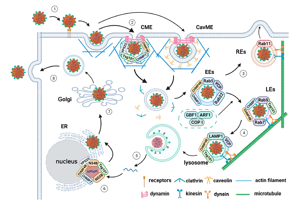
CSFV enters the host through the oronasal mucosa and first infects the crypt epithelium of the tonsils, then spreads systemically through the blood and lymphatics to invade lymphoid tissues. At the cellular level, the interaction of Erns with glycosaminoglycans mediates initial contact, while the E2-E1 heterodimer is required for receptor binding and subsequent endocytosis. Host molecules reported to be involved in CSFV entry include heparan sulfate (HS), the complement regulatory protein CD46, the LDL receptor, integrin β3, annexin II, MERTK, the laminin receptor and ADAM17. Of these, porcine CD46 and HS are considered the main drivers of CSFV attachment and entry.
Innate Immune Evasion
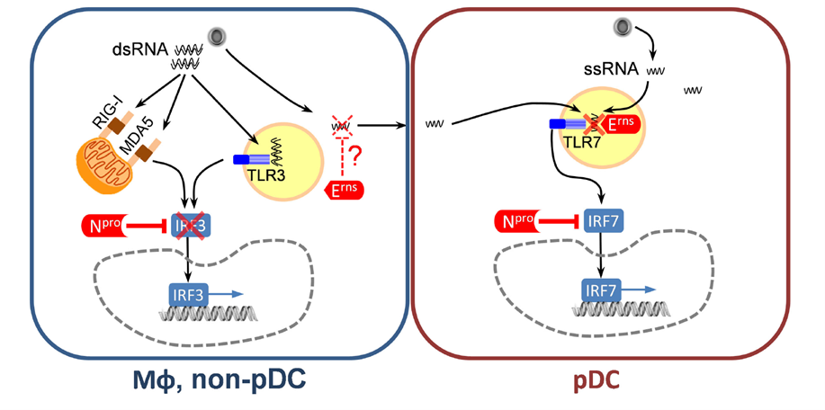
The pathogenicity of CSFV depends largely on its ability to suppress host type I interferon, which it achieves mainly through two proteins, Npro and Erns. Npro interferes with IFN-α/β induction by promoting the degradation of interferon regulatory factor 3 (IRF3). Further studies showed that the Npro–IRF3 interaction not only antagonizes interferon induction but also blocks apoptosis by inhibiting IRF3-dependent translocation of Bax to the mitochondria, allowing infected cells to survive and continue producing virus. Npro can also suppress the expression and nuclear translocation of IRF1, thereby inhibiting type III interferon production. The RNase activity of Erns acts further upstream in immune recognition. Erns degrades viral single-stranded RNA, preventing its recognition by plasmacytoid dendritic cells and thereby reducing interferon production. When the RNase activity of Erns is abolished in a low-virulence strain, humoral immune responses in pigs are enhanced, and the virus's virulence, transmissibility and capacity for persistent infection all decline.
Tissue Damage and Clinical Presentation
CSFV replicates mainly in monocytes/macrophages, dendritic cells and vascular endothelial cells. The disease shows the typical features of a hemorrhagic fever, including thrombocytopenia, platelet aggregation, impaired bone marrow hematopoiesis and generalized lymphoid depletion. Notably, a substantial part of the severe lymphocyte loss results from apoptosis of uninfected bystander cells. Widespread damage to the immune system leads to secondary infections, while endothelial injury and thrombocytopenia together cause hemorrhages in the skin and internal organs. Depending on strain virulence and host status, infection can be acute, chronic or congenital. When pregnant sows are infected with low- or moderate-virulence strains, the virus can cross the placenta to the fetuses, giving rise to immunotolerant piglets that shed virus persistently. These animals are difficult to detect within a herd and pose a major obstacle to eradication.
05Research Targets and Applications
Vaccine Development
Live attenuated vaccines provide earlier and more complete protection than E2 subunit vaccines, but they do not allow infected and vaccinated animals to be distinguished. Using E2 as the vaccine antigen together with Erns antibody testing enables DIVA and is regarded as a promising strategy for the future control and eradication of CSF. The chimeric marker vaccine CP7_E2alf takes a different approach: it uses a BVDV backbone carrying the CSFV E2 gene and has been licensed under the name Suvaxyn CSF Marker. Other strategies, including Npro deletion, inactivation of Erns RNase activity and modification of the TAV epitope, have also been used to design attenuated or marker vaccine candidates.
Diagnostic Development
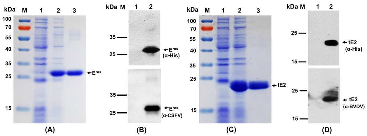
E2 and Erns are the two core antigens for serological testing. Because CSFV and BVDV are serologically cross-reactive, test specificity is a key issue. In one study, a structure-guided truncated CSFV E2 retaining only antigenic domains B/C/D/A was expressed in suspension CHO cells and used to establish an indirect ELISA. The assay showed no cross-reactivity with anti-BVDV sera and could distinguish antibodies against CSFV from those against BVDV. For pathogen detection, RT-PCR usually targets the conserved 5' untranslated region or NS5B, while the E2 gene is commonly used for genotyping.
Antiviral Drugs and Host Targets
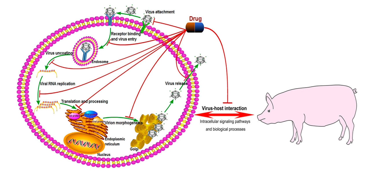
The NS3 protease/helicase and the NS5B polymerase are the main targets for small-molecule antivirals. A class of substituted 2,6-bis(benzimidazol-2-yl)pyridines has been reported to act on an inhibitory hotspot of the pestivirus RdRp. In addition, entry-related host factors such as CD46, HS and ADAM17, as well as host proteins that regulate interferon pathways, offer research directions for non-vaccine control measures.
abinScience CSFV Research Targets
For CSFV diagnostic, vaccine and mechanistic research, abinScience offers recombinant proteins and antibodies against the following targets to support diagnostic assay development, vaccine antigen research and virus–host interaction studies.
- E2: The major immunogen and neutralizing antibody target; suitable for antibody detection, subunit vaccine and receptor-binding studies
- Erns: Involved in viral attachment and immune evasion; a core antigen for DIVA testing
- E1: Forms heterodimers with E2; used in membrane fusion and viral entry studies
- Npro: A key target in immune evasion research, associated with IRF3 degradation and interferon suppression
- p7: A viroporin; useful for studies of virion assembly and release
- NS4A, NS4B, NS5A: Involved in replication complex formation and regulation of RNA replication; useful for studies of viral replication mechanisms
- NS5B: The viral RNA polymerase and a major target for small-molecule antiviral research
- CD46, ADAM17, MERTK: Involved in viral attachment and entry; useful for receptor mechanism studies and exploring non-vaccine control strategies
abinScience CSFV-Related Products
References
- Fan, J.; Liao, Y.; Zhang, M.; Liu, C.; Li, Z.; Li, Y.; Li, X.; Wu, K.; Yi, L.; Ding, H.; et al. Anti-Classical Swine Fever Virus Strategies. Microorganisms 2021, 9, 761. https://doi.org/10.3390/microorganisms9040761
- Li, S.; Wang, J.; Yang, Q.; Naveed Anwar, M.; Yu, S.; Qiu, H.-J. Complex Virus–Host Interactions Involved in the Regulation of Classical Swine Fever Virus Replication: A Minireview. Viruses 2017, 9, 171. https://doi.org/10.3390/v9070171
- Huang, Y.-L.; Meyer, D.; Postel, A.; Tsai, K.-J.; Liu, H.-M.; Yang, C.-H.; Huang, Y.-C.; Berkley, N.; Deng, M.-C.; Wang, F.-I.; et al. Identification of a Common Conformational Epitope on the Glycoprotein E2 of Classical Swine Fever Virus and Border Disease Virus. Viruses 2021, 13, 1655. https://doi.org/10.3390/v13081655
- Summerfield A and Ruggli N (2015) Immune Responses Against Classical Swine Fever Virus: Between Ignorance and Lunacy. Front. Vet. Sci. 2:10. doi: 10.3389/fvets.2015.00010
- Guo, X.; Zhang, M.; Liu, X.; Zhang, Y.; Wang, C.; Guo, Y. Attachment, Entry, and Intracellular Trafficking of Classical Swine Fever Virus. Viruses 2023, 15, 1870. https://doi.org/10.3390/v15091870
- Yi, W., Zhu, H., Wu, Y. et al. The recombinant Erns and truncated E2-based indirect enzyme-linked immunosorbent assays to distinguishably test specific antibodies against classical swine fever virus and bovine viral diarrhea virus. Virol J 19, 121 (2022). https://doi.org/10.1186/s12985-022-01851-w


 中文
 English
 한국어
 日本語
 Español
 Français
 Русский