
 中文
中文 English
 한국어
한국어 日本語
日本語 Español
 Français
 Русский


| Catalog No. | HB058016 |
|---|---|
| Description |
Tisotumab (HB058016) is a research-grade recombinant antibody targeting CD142. Produced in mammalian cells with native-like glycosylation.
Highlights
|
| Species reactivity | Human |
| Applications | ELISA, Bioactivity: FACS, Functional assay, Research in vivo |
| Host species | Human |
| Isotype | IgG1-kappa |
| Expression system | Mammalian cells |
| Clonality | Monoclonal |
| Tested applications | WB, FCM |
| Target | Tissue factor, CD142, F3, Thromboplastin, Coagulation factor III, TF |
| Endotoxin level | < 10 EU/mg |
| Purity | >95% purity as determined by SDS-PAGE. |
| Purification | Protein A/G purified from cell culture supernatant. |
| Accession | P13726 |
| Form | Liquid |
| Storage buffer | 0.01M PBS pH 7.4 Please refer to the specific buffer information in the hardcopy of datasheet or the lot-specific COA. |
| Stability and Storage | Use a manual defrost freezer and avoid repeated freeze thaw cycles. Store at 2 to 8°C for frequent use. Store at -20 to -80°C for twelve months from the date of receipt. |
| Alternate Names | HuMax-TF-ADC, TF-011-MMAE, HuMax-TF, 1418628-81-5 |
| Background | Tissue factor (CD142/F3) is a ~33 kDa protein. Initiates blood coagulation by forming a complex with circulating factor VII or VIIa. The [TF:VIIa] complex activates factors IX or X by specific limited proteolysis. TF plays a role in normal hemostasis by initiating the cell-surface assembly and propagation of the coagulation protease cascade. CD142 is the therapeutic target of tisotumab vedotin (Tivdak). |
| Note | For research use only. Not suitable for clinical or therapeutic use. |
SEC-HPLC detection for Research Grade Tisotumab.
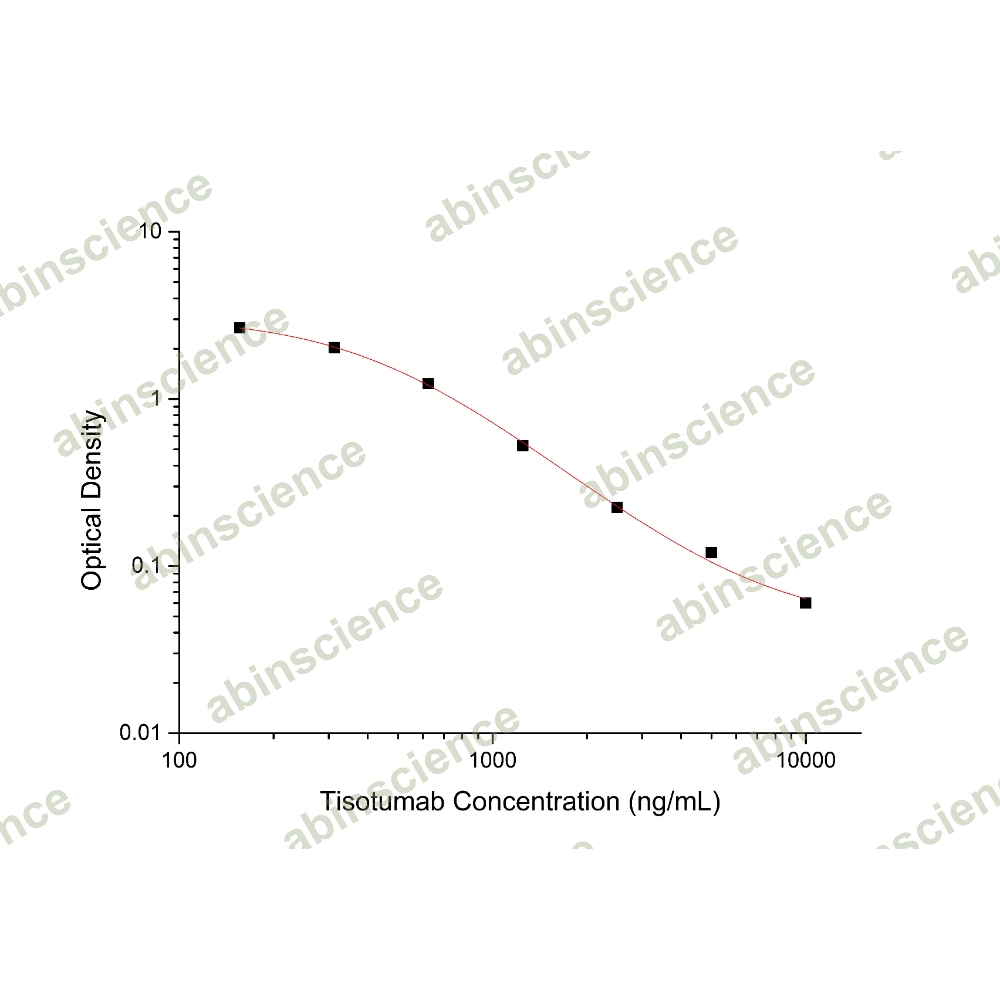
Detects CD142/F3/TF in indirect ELISAs.
SDS-PAGE for Research Grade Tisotumab
Flow-cytometry using anti-human CD142 antibody. Untransfected cells (blue Histogram) and Transfected cells (Yellow Histogram) were stained with an anti-human CD142 monoclonal antibody (Catalog: HB058016) at a concentration of 5 µg/ml for 30 mins at RT. After washing, bound antibody was detected using a Goat Anti-Human IgG H&L Polyclonal Antibody, FITC (abinScience: HF690414) and cells analysed on a NovoCyte Flow Cytometer.
Western blot analysis was performed using anti-CD142/F3/TF monoclonal antibody at 1μg/ml on various samples.
Lane 1: recombinant human CD142/F3/TF (Catalog No: HB058011), 50ng
Lane 2: recombinant human CD142/F3/TF (Catalog No: HB058011), 30ng
Lane 3: recombinant human CD142/F3/TF (Catalog No: HB058011), 20ng
Lane 4: recombinant human CD142/F3/TF (Catalog No: HB058011), 10ng
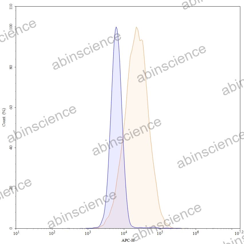
Flow-cytometry using APC anti-human CD142 antibody. A431 cells were stained with an irrelevant antibody (Blue Histogram) or an APC anti-human CD142 monoclonal antibody (Catalog HB058016, Yellow Histogram) at a concentration of 5 µg/ml for 30 mins at RT. After washing, and cells analysed on a NovoCyte Flow Cytometer.
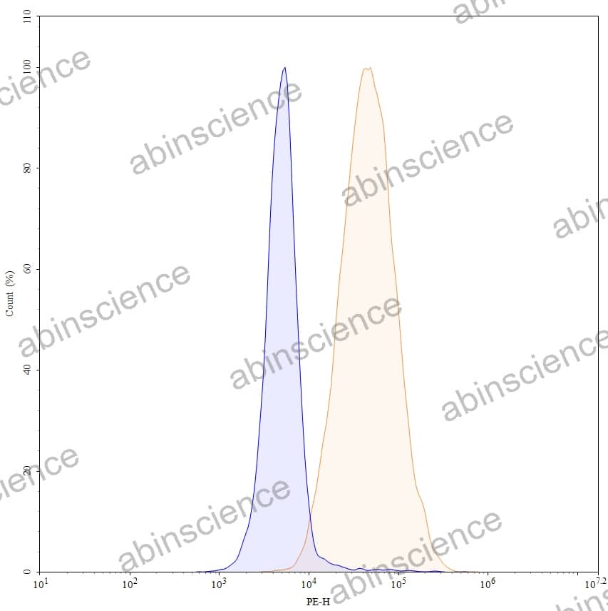
Flow-cytometry using PE anti-human CD142 antibody. A431 cells were stained with an irrelevant antibody (Blue Histogram) or an PE anti-human CD142 monoclonal antibody (Catalog HB058016, Yellow Histogram) at a concentration of 5 µg/ml for 30 mins at RT. After washing, and cells analysed on a NovoCyte Flow Cytometer.
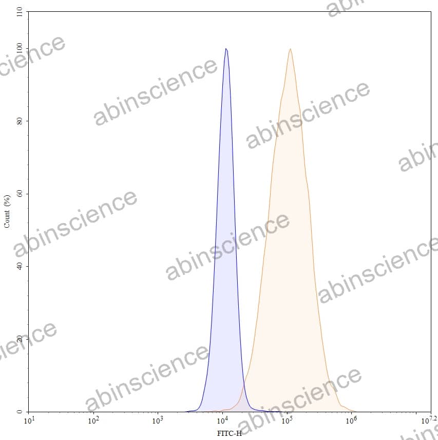
Flow-cytometry using FITC anti-human CD142 antibody. A431 cells were stained with an irrelevant antibody (Blue Histogram) or an FITC anti-human CD142 monoclonal antibody (Catalog HB058016, Yellow Histogram) at a concentration of 5 µg/ml for 30 mins at RT. After washing, and cells analysed on a NovoCyte Flow Cytometer.


Contact us for custom quotes, bulk requests and any other issues.
Mail: support@abinScience.com
+86-27-65523339
Building C, No. 666, Shen Dun Si Lu, Wuhan, 430206, China


