
 中文
中文 English
 한국어
한국어 日本語
日本語 Español
 Français
 Русский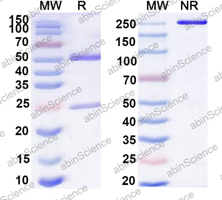
| Catalog No. | VK683043 | ||||
|---|---|---|---|---|---|
| Species reactivity | Adeno-associated virus 2 (AAV2) | ||||
| Applications | Dot blot, ELISA, ICC/IF, IP, Neutralization | ||||
| Host species | Mouse | ||||
| Clonality | Monoclonal | ||||
| Isotype | IgG3 | ||||
| Clone ID | A20 | ||||
| Target | Capsid protein VP1, VP1 | ||||
| Endotoxin level | <10 EU/mg | ||||
| Purity | >95% as determined by SDS-PAGE. | ||||
| Purification | Protein A/G purified from cell culture supernatant. | ||||
| Accession | P03135 | ||||
| Form | Liquid | ||||
| Storage buffer | 0.01M PBS pH 7.4 Please refer to the specific buffer information in the hardcopy of datasheet or the lot-specific COA. |
||||
| Product Usage Information |
| ||||
| Background | Capsid protein VP1 is a ~81 kDa protein. Capsid protein self-assembles to form an icosahedral capsid with a T=1 symmetry, about 22 nm in diameter, and consisting of 60 copies of three size variants of the capsid protein VP1, VP2 and VP3 which differ in their N-terminus. The capsid encapsulates the genomic ssDNA. Binds to host cell heparan sulfate and uses host ITGA5-ITGB1 as coreceptor on the cell surface to provide virion attachment to target cell. This attachment induces virion internalization predominantly through clathrin-dependent endocytosis. Binding to the host receptor also induces capsid rearrangements leading to surface exposure of VP1 N-terminus, specifically its phospholipase A2-like region and putative nuclear localization signal(s). | ||||
| Note | For research use only |
SDS-PAGE for Anti-AAV2 Capsids (intact particle) Antibody (A20)


Contact us for custom quotes, bulk requests and any other issues.
Mail: [email protected]
+86-27-65523339
Building C, No. 666, Shen Dun Si Lu, Wuhan, 430206, China


