Please ensure Javascript is enabled for purposes of website accessibility
Home > Featured Products > Bacteria & Viruses-related products > Nipah Virus (NiV) / Hendra Virus (HeV)

Nipah Virus (NiV) / Hendra Virus (HeV) Research Reagents

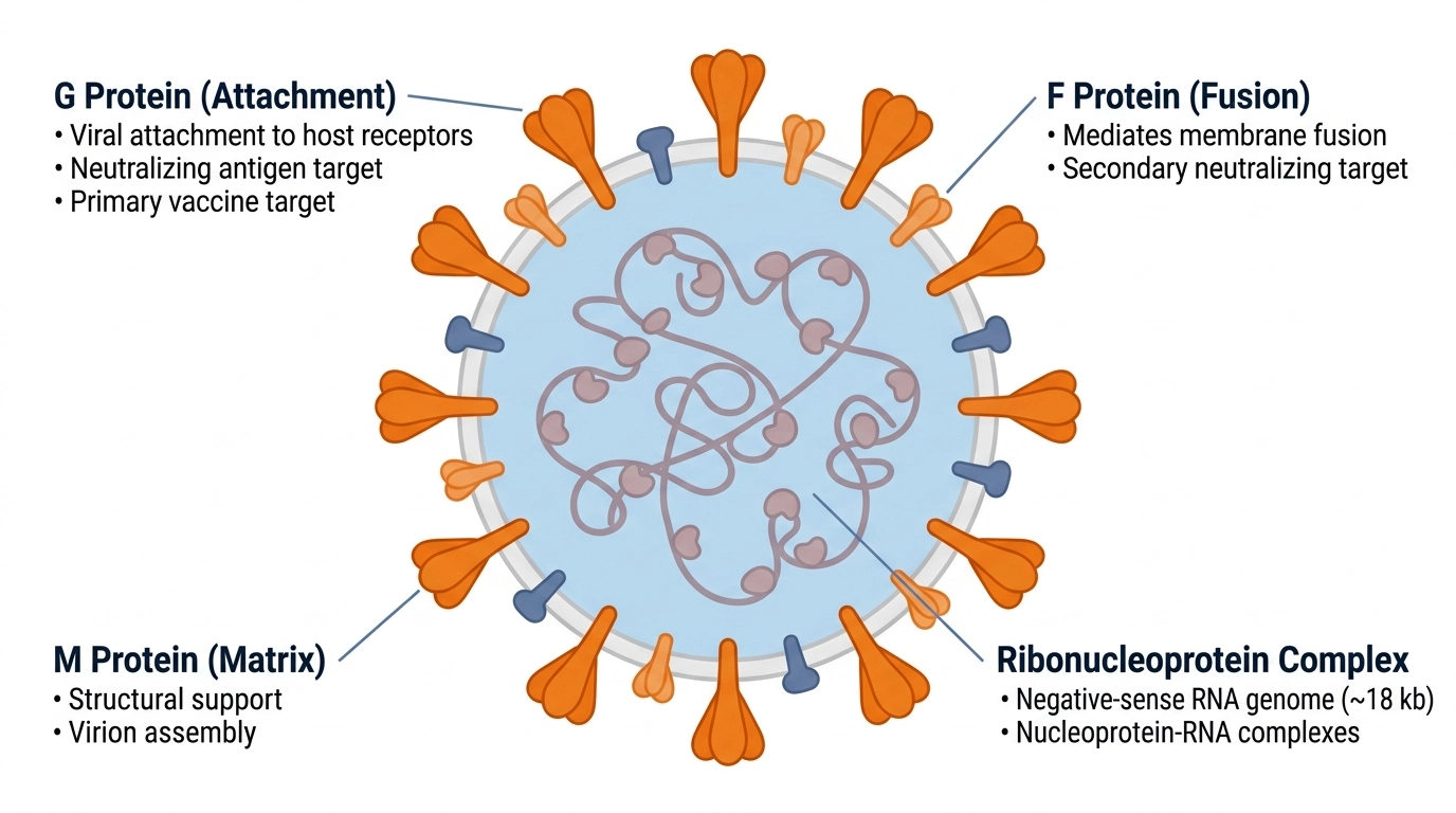
Nipah virus (NiV) and Hendra virus (HeV) are enveloped, negative-sense single-stranded RNA viruses of the genus Henipavirus, family Paramyxoviridae, with case fatality rates of 40-75% in humans. Both viruses are zoonotic pathogens transmitted from bat reservoirs to intermediate hosts and humans, causing severe respiratory and neurological disease. The virion is composed of two surface glycoproteins: G (attachment) and F (fusion), together with matrix protein M and ribonucleoprotein complex. G and F are primary targets for neutralizing antibodies and vaccines. NiV/HeV-specific antibodies, recombinant proteins, and assay kits enable vaccine development, therapeutic antibody discovery, seroprevalence studies, and biosafety diagnostics.

Research Use Only (RUO)BSL-4 pathogen research applications.

Nipah/Hendra virus structure — G and F glycoproteins, matrix protein

Fig. 1 Nipah/Hendra virus structure. G protein (attachment) and F protein (fusion) are the primary surface glycoproteins; matrix protein (M) and ribonucleoprotein complex form the virion core.

abinScience provides neutralizing monoclonal antibodies, InVivoMAb-grade antibodies, and recombinant G/F glycoprotein antigens for Nipah/Hendra vaccine and therapeutic development. Products are manufactured by AtaGenix Laboratories under ISO 9001 & ISO 13485. Contact us for neutralization assays and BSL-4 diagnostic optimization.

Key Research Targets

G Protein (Attachment Glycoprotein) — G mediates viral attachment to host cell receptors and is the primary target for neutralizing antibodies and vaccines. G-specific antibodies (mAb m102.3, VK685013; pAb VK631014; mAbs VK685015, VK685025, VK685035) block viral entry and are highly protective. Recombinant G protein serves as ELISA coating antigen, immunogen, and vaccine candidate substrate. Essential for neutralization assays and passive immunization studies.
→ Browse G Protein products

F Protein (Fusion Glycoprotein) — F mediates viral membrane fusion and is a secondary neutralizing antigen target. F-specific polyclonal antibodies (VK521014) enable fusion inhibition assays and vaccine immunogenicity assessment. F antigen serves as vaccine component and diagnostic coating antigen. Fusion-inhibiting antibodies support therapeutic development.
→ Browse F Protein products

N & M Proteins (Nucleoprotein & Matrix) — N forms ribonucleoprotein complex (VK432014); M provides structural support (VK408014, VK499014). Used for diagnostic antigen detection, viral confirmation assays, and seroprevalence studies. Non-glycoprotein targets for assay multiplexing.
→ Browse N/M Protein products

Recommended Applications

Vaccine Development & Potency Testing — Recombinant G/F antigens are vaccine immunogens. G/F-specific antibodies quantify neutralizing responses and predict vaccine efficacy in animal models and clinical trials.

Therapeutic Antibody Development — Neutralizing monoclonal antibodies (m102.3, 1A050, 1A206, 1A207) serve as reference standards for therapeutic drug discovery. InVivoMAb-grade antibodies enable in vivo neutralization and passive immunization studies.

Diagnostic & Biosafety Assays — G/F recombinant antigens enable rapid diagnostic ELISA and lateral flow assay development. N/M antigens support confirmatory multiplex assays for clinical and field diagnostics.

Seroprevalence & Epidemiology — G-specific ELISA kits enable population serosurveillance in high-risk regions (Bangladesh, Malaysia, Australia) and bat reservoir screening.

References

1. Luby SJ. The pandemic potential of Nipah virus. Antiviral Res. 2013;100(1):38-43.

2. Clayton AJ. The comparative biology of Nipah and Hendra viruses. Curr Opin Virol. 2016;16:99-105.

3. Eaton BT, Broder CC, Middleton D, et al. Hendra and Nipah viruses: different and dangerous. Nat Rev Microbiol. 2006;4(1):23-35.

4. Rockx B, Corti D, Donaldson E, et al. Structural basis of human antibody-mediated neutralization of Nipah virus. Nat Microbiol. 2018;3(2):143-150.

5. Long JS, Honko A, Basler CF. Interferon and interferon-induced protein expression in respiratory epithelial cells during Henipavirus infection. Virology. 2014;468-470:54-62.

All products manufactured by AtaGenix Laboratories under ISO 9001 & ISO 13485. Contact us for custom antibody development and BSL-4 research applications.

57 product results for "Nipah Virus (NiV) / Hendra Virus (HeV)"

Options+
Options
Confirm
  • Product types
    0 selected
    Antibodies (45)
    Proteins (10)
    Assay Kit (2)
  • Expression system
    0 selected
    E. coli (7)
    Mammalian cells (3)
  • Target
    0 selected
    Fusion glycoprotein F1 (1)
    Fusion glycoprotein F0 (10)
    Prefusion Protein (2)
    Fusion Protein (1)
    Fusion glycoprotein/F Trimer (1)
    Glycoprotein G (30)
    M/Protein M (2)
    Fusion glycoprotein F2 (2)
    M/Matrix protein (2)
    F/Fusion glycoprotein F0 (3)
    Protein N/Nucleoprotein (3)
  • Species
    0 selected
    Hendra virus (HeV) (3)
    Nipah virus (NiV) (22)
  • Applications
    0 selected
    Bioactivity testing in progress (10)
    FCM (3)
    SPR (2)
    ELISA (48)
    Neutralization (27)
    WB (22)
    SDS-PAGE (10)
    IP (3)
    IHC (5)
    Immunogen (10)
  • Reactivity
    0 selected
    Nipah virus (10)
    Hendra virus (HeV) (13)
    Hendra virus (7)
    Nipah virus (NiV) (20)
  • Host species
    0 selected
    Alpaca (1)
    Rabbit (5)
    Mouse (9)
    Human (26)
  • Clonality
    0 selected
    Polyclonal (5)
    Monoclonal (40)
  • Conjugate
    0 selected
    Unconjugated (40)